TAMRA NHS ester
TAMRA NHS ester
Cat. No. List below
Description
Key features:
1. Bright yellow fluorescence with excellent photostability.
2. Excitation/Emission Maxima: 554/583 nm.
3. Extinction Coefficient: ≥ 68,000 cm?¹M?¹.
4. CF280: 0.3.
5. Molecular Weight: 529.55 g/mol.
6. Appearance: Red solid.
7. Solubility: DMF, DMSO.
8. Storage Conditions: -20°C, protected from light.
Applications:
1. Labeling of antibodies, peptides, proteins, and oligonucleotides.
2. Automated DNA sequencing.
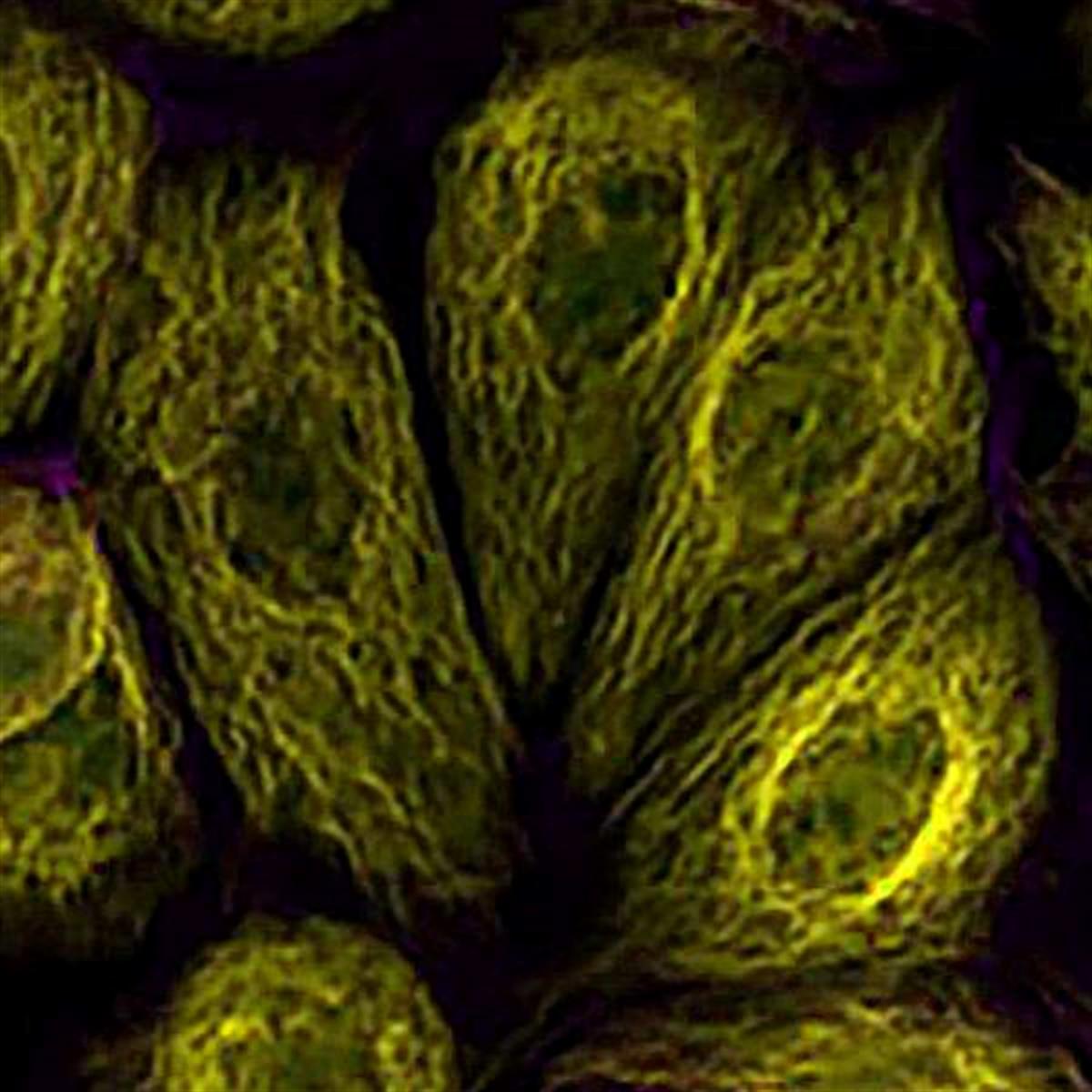
3. Fluorescence microscopy and confocal imaging.
4. Flow cytometry for cell analysis.
5. FRET (Fluorescence Resonance Energy Transfer) studies as an acceptor for FAM fluorophore.
6. Molecular tracking and localization in biological systems.
Advantages:
1. High Reactivity: The NHS ester group reacts efficiently with primary amines (e.g., ε-amino groups of lysine residues or the amine terminus of nucleotides), forming stable amide bonds.
2. Wide Compatibility: Suitable for labeling proteins, peptides, and oligonucleotides in various experimental setups.
3. Excellent Optical Properties: Bright fluorescence with high extinction coefficient ensures sensitivity in detection.
4. Versatility: Can be used in diverse applications such as DNA sequencing, protein conjugation, and live-cell imaging.
5. Bioorthogonality: Minimal interference with native biochemical processes during labeling.
TAMRA NHS ester is particularly well-suited for preparing fluorescent bioconjugates such as labeled antibodies or avidin derivatives. It is also widely utilized in oligonucleotide labeling for automated DNA sequencing applications. Its compatibility with common laser lines (543 or 546 nm) and its spectral similarity to other popular dyes like Cy3 make it an excellent choice for researchers looking to integrate it into existing workflows.
- Fluorophore: TAMRA
- Reactive group: NHS ester
- Excitation/Emission Max.(nm): 554/583
- Spectrally similar dyes: DyLight549, Cy3, ATTO550
- Extinction coefficient: ≥ 68,000 cm-1M-1
- CF280: 0.3
- Appearance: Red Solid
- Molecular Weight: 529.55 g/mol
- Solubility: DMF, DMSO
- Storage conditions: -20 ℃, protect from light
| Quick link (Cat.#) | Series | Quick link (Cat.#) | Series |
| KWS1025 | TAMRA NHS ester | KWSN1025 | TAMRA Sulfo-NHS ester |
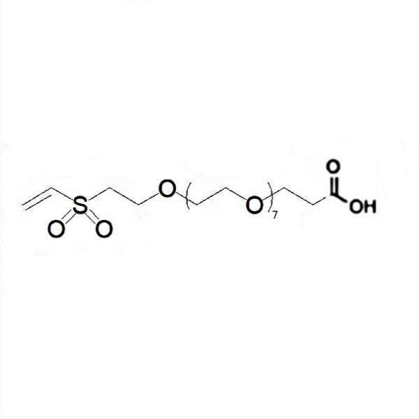
| KWA1020 | TAMRA Vinylsulfone | KWM1057 | TAMRA Maleimide |
| KWZ1025 | TAMRA Azide | KWH1025 | TAMRA Hydrazide |
| KWK1025 | TAMRA Alkyne | KWG1025 | TAMRA PEG4-Alkyne |
| DWR1001 | TAMRA ADIBO | KWE1025 | TAMRA Amine |
| KWT1057 | TAMRA Thiol | KWR2025 | TAMRA Dichlorotriazine |
Background
Other Labeling Dyes
BioActs provides other traditionally used dyes such as 5(6)-Carboxyfluorescein (FAM) and 5(6)-Carboxytetramethylrhodamine (TAMRA) dyes for labeling of biomolecules. FAM is one of popular green fluorescent reagents used for labeling peptides, proteins and nucleotides. In addition to relatively high absorptivity, good fluorescence quantum yield and good water solubility, FAM has an excitation maximum that closely matches the 488 nm spectral line of the argon-ion laser. TAMRA fluorophore has been a widely used for preparing bioconjugates, especially fluorescent antibody and avidin derivatives. TAMRA dye is also widely utilized for oligonucleotide labeling and automated DNA sequencing applications. TAMRA is often used as FRET acceptor for FAM fluorophore.
- Other fluorescent dyes such as Cyanine, ICG, TAMRA, FAM, etc. are also available.
- All dyes are equipped with various reactive and functional groups.
- High quality and excellent performance
Table 1. TAMRA dye applications


Figure 1. Structure of FAM and TAMRA dyes
Citation & Reference
1. PYARE L. KHANNAA. 4',5'-Dimethoxy-6-carboxyfluorescein: a novel dipole-dipole coupled fluorescence energy transfer acceptor useful for fluorescence immunoassays. Anal Biochem 108.1 (1980): 156-61.
2. Torimura M. Fluorescence-quenching phenomenon by photoinduced electron transfer between a fluorescent dye and a nucleotide base. Anal Sci 17.1 (2001): 155-60.
3. Sylvie Soulie-Begu. In-vivo pharmacokinetic study of two fluorescein derivatives by fluorescence spectroscopy. Optical Biopsies 2627 (1995).
4. Danny van Lierop. Positively charged silver nanoparticles and their effect on Surfaceenhanced Raman scattering of dye-labelled oligonucleotides. Chem. Commun 48 (2012): 8192-8194.
5. Pete Theisen. Fluorescent Dye Phosphoramidite Labelling of Oligonucleotides. Tetrahedron Letters 33.35 (1992): 5033-5036.