Anti-Rad18 (mouse) antibody, rabbit polyclonal IgG
Product#: FNK-70-025
$346.79
Availability:
Ships in 1-2 Weeks
Anti-Rad18 (mouse) antibody, rabbit polyclonal IgG
General information
| Cat. No. | :FNK-70-025 |
| Size | :100 ug |
| Antigen | :Mouse |
| Host Species | :Rabbit |
| Reactivity | :Mouse Rad18 protein. Not reactive to human Rad18. |
| Immunogen | :GST-fusion protein containing 100 carboxyl terminal amino acids of mouse Rad18 |
| Product | :IgG fraction of anti-mouse Rad18 rabbit serum |
| Application | :1) Western blotting (1,000 fold dilution) :2) Immuno-precipitation (200~500 dilution)) :3) Indirect immuno-fluorescence staining. (assay dependent) :4) Immuno-histochemistry (100~300 fold dilution) |
| Storage | :Ship at 4℃. Upon arrival, spin-down and store at -20℃ |
| Form | :1 mg/ml in PBS, 50% glycerol, filter-sterilized. Azide and carrier-protein free |
| Data Link | :UniProtKB/Swiss-Prot Q9QXK2 Mouse Rad18 Gene ID 16098139 Mouse Rad18 |
Description
The Rad6 (UBE2B)-Rad18 pair of genes plays a critical role in post-replication repair of damaged DNA. Rad6 protein functions as an E2 enzyme and Rad18 (509 aa, 57.4 kDa) as a ubiquitine ligase (E3) which ubiquitinates PCNA. Rad18 recruits translesion DNA polymerases to damaged DNA.
 |
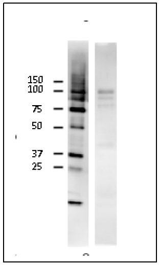 |
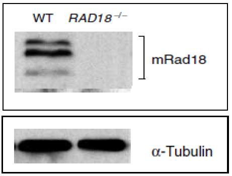
Fig.1.Identification of mouse Rad18 protein in ES cells by Western blot with anti-mRad18 antibody. WT; Lysate of wild-type mouse ES cells RAD18-/-; Lysate of Rad18 double knock-out mouse ES cells Protein levels of α -tubulin in the lysates are shown as a control. Three bands are absent in RAD18 knock-out cells. |
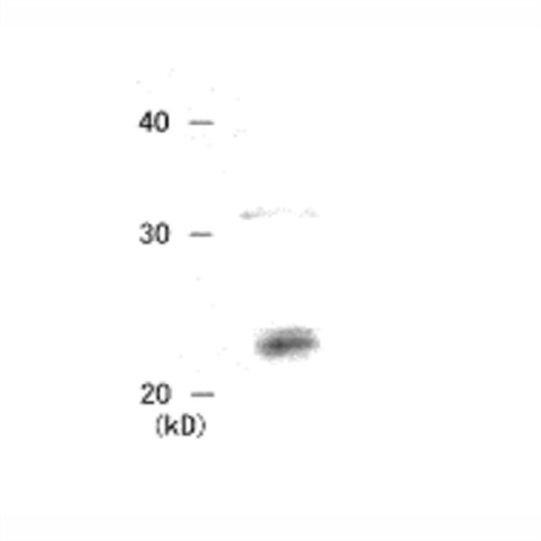
Fig.2. Identification of mouse Rad18 protein in NIH3T3 cells by western blot with the antibody.
Cell extract, 23 ug,was used. Anti-mouse Rad18 was used at 2,000 fold dilution. Similar to Fig.1, two extra bands (75~90 kDa) may represent modified products |
 |
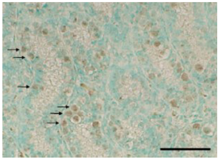 |
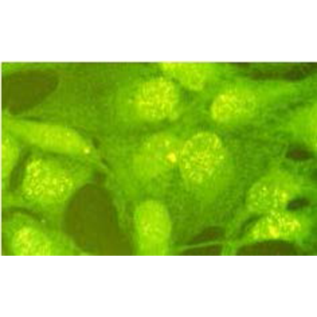
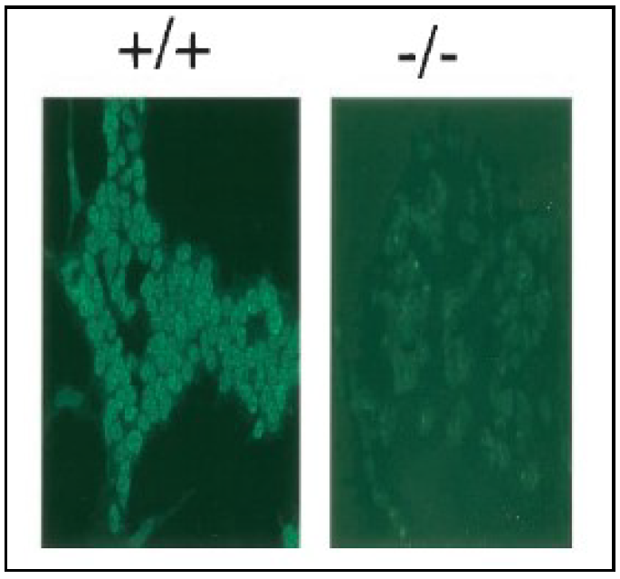
Fig.3. Immunofluorescence staining of Rad18 protein with anti-mRad18 antibody. Wild-type (+/+) and RAD18-/- ES cells (-/-). Samples were prefixed 3.7% formaldehyde and fixed with 80% methanol. Anti-mRad18 antibody was used at 1/300 dilution. AS a second antibody,FITC conjugated anti-rabbit IgG was used |
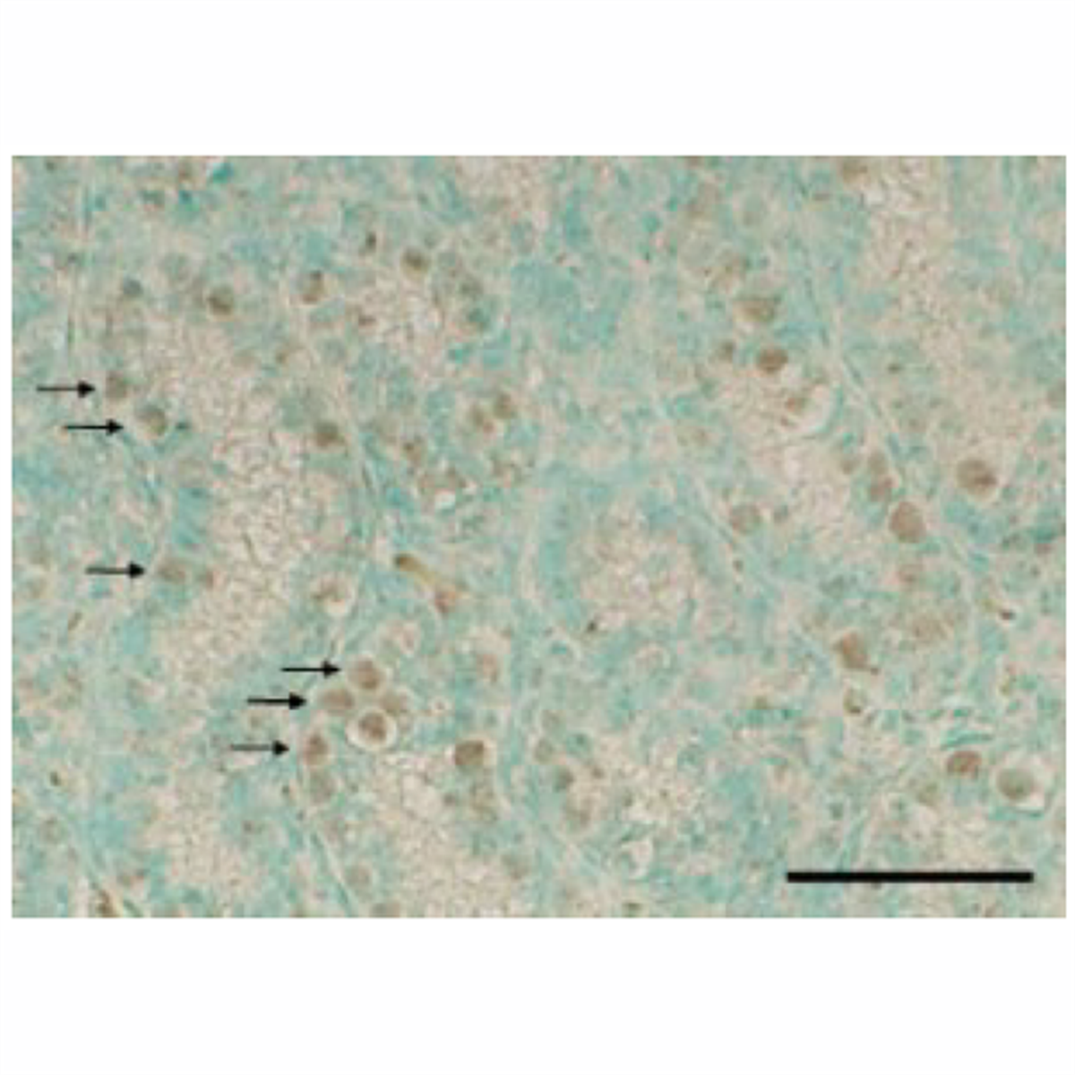
Fig.4. Detection of Rad18 in mouse testis.
Section of paraformaldehyde fixed mouse was stained with anti-mRad18 antibody. As a second antibody, peroxidase-conjugated anti-rabbit IgG donky antibody was used. Signals were enhanced with TSA plus biotin system and detected by using DAB substrate. Arrows indicate undifferentiated spermatogonia.
|
References
- Tateishi S. et al. (2003) Enhanced genomic instability and defective postreplication repair in RAD18 knockout mouse embryonic stem cells. Mol Cell Biol 23:474-81. PubMed 12509447 WB, IF/IC
- Watanabe K. et al. (2004) Rad18 guides poleta to replication stalling sites through physical interaction and PCNA monoubiquitination. EMBO J. 23:3886-96. PubMed 15359278 WB
- Masuyama S. et al. (2005) Regulated expression and dynamic changes in subnuclear localization of mammalian Rad18 under normal and genotoxic conditions. Genes Cells. 10:753-62. PubMed 16098139 IHC
- Sun J. et al. (2009) Rad18 is required for long-term maintenance of spermatogenesis in mouse testes. Mech Dev 126:173-83.PubMed 19068231 IHC, WB
Aliases for RAD18 Gene
- RAD18 E3 Ubiquitin Protein Ligase 2 3 5
- RNF73 2 3 4
- RING-Type E3 Ubiquitin Transferase RAD18 3 4
- Postreplication Repair Protein RAD18 3 4
- E3 Ubiquitin-Protein Ligase RAD18 3 4
- RING Finger Protein 73 3 4
- HRAD18 3 4
- HHR18 3 4
- Postreplication Repair Protein HRAD18p 3
- RAD18 Homolog (S. Cerevisiae) 2
- RAD18, S. Cerevisiae, Homolog 3
- RAD18 Homolog 3
- EC 2.3.2.27 4
- RAD18 5