Hyp-Stamp, H2O2-Responsive Protein Labeling Reagent
Product#: FNK-FDV-0052
$538.20
Availability:
Ships in 48 hours
Hyp-Stamp, H2O2-Responsive Protein Labeling Reagentv
Cat. No.: FNK-FDV-0052
Description
Hyp-Stamp is an innovative H2O2-responsive protein labeling reagent designed for visualizing and analyzing cellular hydrogen peroxide localization. This advanced fluorescent probe offers unique capabilities for studying oxidative stress and redox signaling in biological systems.
Key Features:
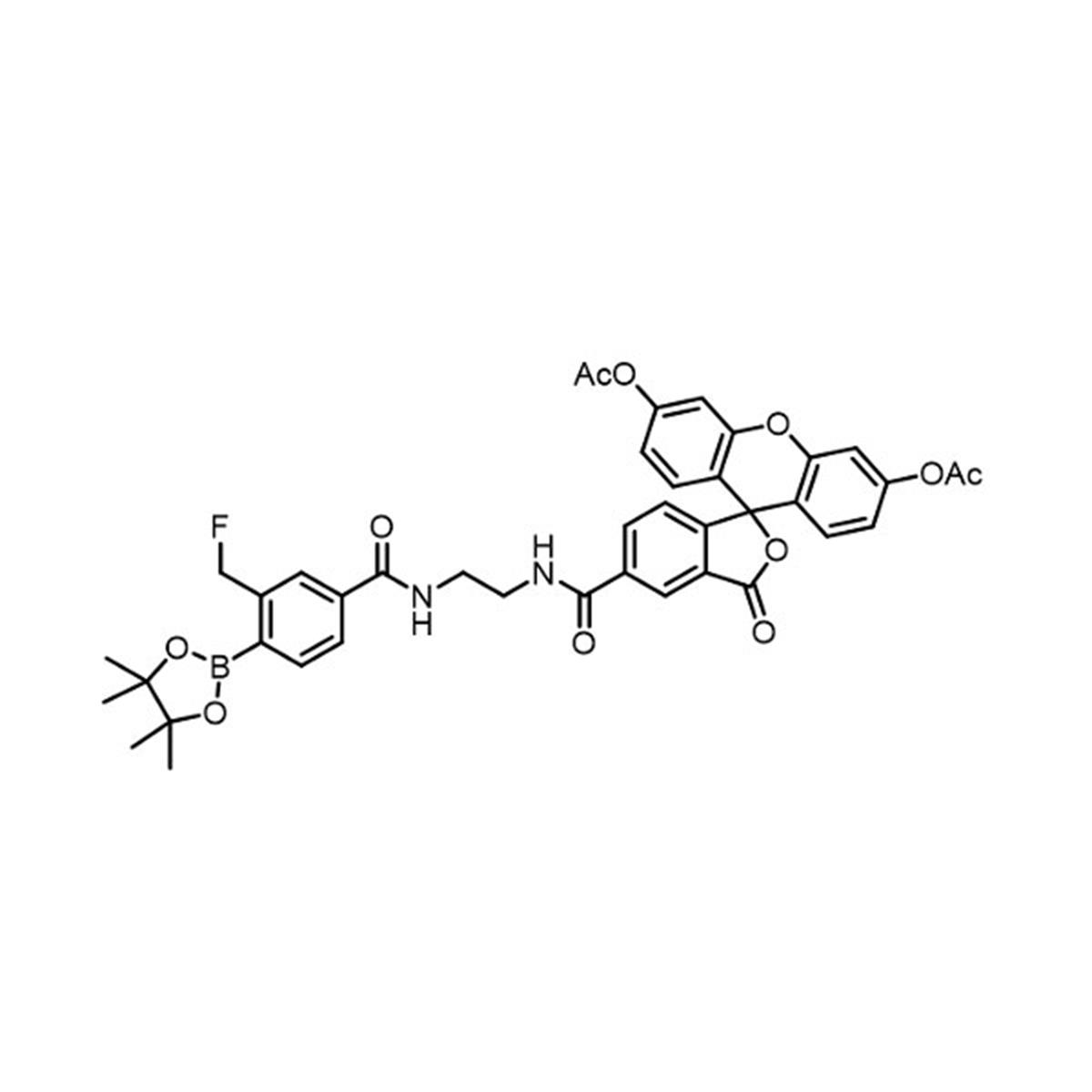
1. Molecular formula: C41H38BFN2O11
2. Molecular weight: 764.56 g/mol
3.Excitation/Emission: 495 nm/515 nm
4. Compatible with FITC filter sets
5. Cell-permeable due to diacetylfluorescein (FDA) moiety
6. Activates upon cleavage by intracellular esterases
7. H2O2-responsive protein labeling
Applications:
1. Visualization of H2O2 localization in fixed cells and tissues
2. Fluorescent detection in SDS-PAGE
3. Western blotting following anti-fluorescein immunoprecipitation
4. Proteomic analysis via LC-MS/MS after immunoprecipitation
Advantages:
1. Specific H2O2 detection: Responds selectively to cellular H2O2, enabling precise localization studies
2. Retained signal after fixation: Unlike conventional probes, allows visualization in fixed samples
3. Versatile analysis options: Compatible with various protein analysis techniques
4. Cell-permeable design: Efficiently enters cells for intracellular labeling
5. Bright fluorescence: Strong green emission for sensitive detection
6. Multimodal imaging: Suitable for both microscopy and biochemical assays
Important Notes:
1. Exhibits no fluorescence until cleaved by intracellular esterases
2. Prepare aqueous solutions immediately before use due to potential hydrolysis
3. Avoid storing aqueous solutions
Hyp-Stamp offers researchers a powerful tool for studying H2O2-mediated cellular processes, combining the specificity of H2O2 detection with the versatility of protein labeling. Its unique properties enable detailed investigations into oxidative stress and redox signaling pathways across various experimental platforms.
Specification
- Formulation: C41H38BFN2O11
- Molecular weight: 764.56 g/mol
- Solubility: Soluble in DMSO
- Ex/Em: 495 nm/515 nm
- *FITC filter sets are available.
- Size: 100 µg
- Storage: -20°C
Note: This reagent contains a diacetylfluorescein (FDA), which promotes cell-permeability and exhibits no fluorescence. A strong green fluorescence emission occurs when the FDA is cleaved by intracellular esterases and converted to fluorescein. Hyp-Stamp can be slowly hydrolyzed in an aqueous medium. Thus, we recommend to prepare the Hyp-Stamp aqueous solution, use it immediately, and avoid storing it.
Reconstitution and StorageReconstitution: Stock solution in 100% DMSO.
Storage: Store powder at -20oC.
After reconstitution in DMSO, make aliquots and store at -20 °C.
Avoid repeated freeze-thaw cycles. Protect from light.
How to use
General procedure for cellular H2O2 imaging
1. Culture cells on appropriate dishes.
2. Remove the culture medium and wash cells several times.
3. Stimulate cells with any reagents such as oxidative stress inducer or immune stimulant and incubate cells for the appropriate time.
4. Incubate cells with 5 µM of Hyp-Stamp diluted in serum-free medium for 30 min. NOTE: Empirically optimize and determine the concentration of Hyp-Stamp for your experiments.
5. The cells are fixed with chilled methanol or formaldehyde solution, washed with PBS, and imaged by fluorescence microscope.
NOTE: You may need to optimize cell fixation conditions, including selecting a fixative solution, time, and temperature for your experiments.
Western blotting of Hyp-Stamp treated cell lysate
2. Remove the culture medium and wash cells several times.
3. Stimulate cells with any reagents such as oxidative stress inducer or immune stimulant and incubate cells for the appropriate time.
4. Incubate cells with 5 µM of Hyp-Stamp diluted in serum-free medium for 30 min. NOTE: Empirically optimize and determine the concentration of Hyp-Stamp for your experiments.
5. The cells are fixed with chilled methanol or formaldehyde solution, washed with PBS, and imaged by fluorescence microscope.
NOTE: You may need to optimize cell fixation conditions, including selecting a fixative solution, time, and temperature for your experiments.
1. Stimulate cells with any reagent and treat with Hyp-Stamp, as described above.
2. The treated cells are washed several times with PBS and lysed in lysis buffer, such as RIPA buffer, containing a protease inhibitor cocktail.
3. Apply the samples to SDS-PAGE and electrotransfer onto PVDF membranes.
4. Treat the membranes with anti-fluorescein antibodies to detect fluorescein-labeled proteins.
2. The treated cells are washed several times with PBS and lysed in lysis buffer, such as RIPA buffer, containing a protease inhibitor cocktail.
3. Apply the samples to SDS-PAGE and electrotransfer onto PVDF membranes.
4. Treat the membranes with anti-fluorescein antibodies to detect fluorescein-labeled proteins.
Preparation of the lysate of Hyp-Stamp treated cells and enrichment of fluorescein-labeled proteins
1. Stimulate cells with any reagent and treat with Hyp-Stamp, as described above.
2. The treated cells are washed several times with PBS and lysed in lysis buffer, such as RIPA buffer, containing a protease inhibitor cocktail.
3. Centrifuge the lysates and collect the supernatant.
4. Small molecules, including an excess Hyp-Stamp, are removed from the lysates with any appropriate process (i.e., dialysis, ultrafiltration, and protein extraction).
5. The resulting protein solutions are immunoprecipitated with anti-fluorescein antibodies.
2. The treated cells are washed several times with PBS and lysed in lysis buffer, such as RIPA buffer, containing a protease inhibitor cocktail.
3. Centrifuge the lysates and collect the supernatant.
4. Small molecules, including an excess Hyp-Stamp, are removed from the lysates with any appropriate process (i.e., dialysis, ultrafiltration, and protein extraction).
5. The resulting protein solutions are immunoprecipitated with anti-fluorescein antibodies.
Reference data
Ractivity of the protein-binding moiety of Hyp-Stamp with several reactive oxygen species (ROS) and reactive nitrogen species (RNS)
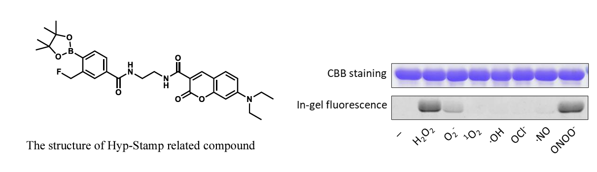
200 µM of several ROS and RNS were added to a PBS (−) solution of BSA (10 µM) containing 50 µM of HypStamp related compound having the same protein-binding moiety as Hyp-Stamp (see structure below), and the mixture was incubated for 30 min. The samples were applied to a 12.5% SDS–PAGE gel and imaged by Coomassie Brilliant Blue (CBB) and in-gel fluorescence. As a result, the protein binding moiety was found to selectively react with H2O2 compared with other reactive species, except for peroxynitrite (ONOO–), which was also able to trigger the BSA labeling. In general, the cellular concentration of ONOO- is much lower than H2O2. Nevertheless, the control experiments using ONOO- generation inhibitor such as NG-nitro-L-arginine methyl ester (L-NAME) are recommended to determine whether the specific analysis of H2O2 by Hyp-Stamp with eliminating the influence of ONOO-.
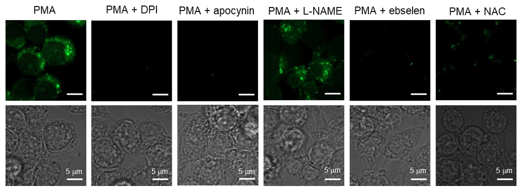
Imaging of the H2O2 localization by immunostimulation in macrophage cells
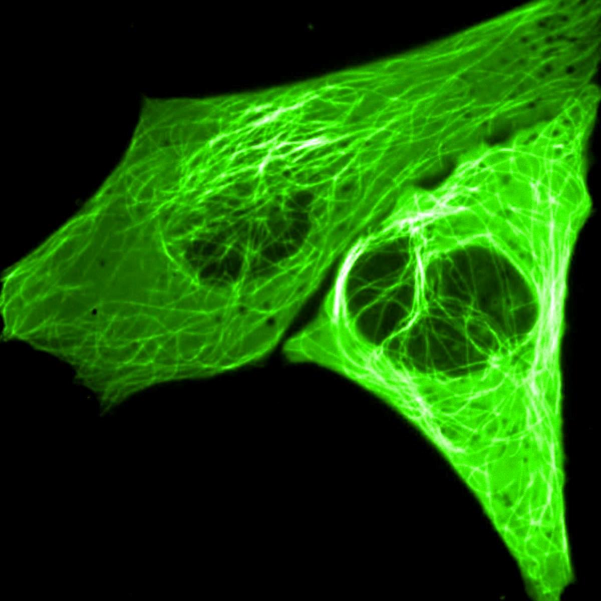
RAW264.7 cells (murine macrophage cell line) were immunostimulated (treated with 1 µg/mL PMA) in the presence of various inhibitors or scavengers of reactive species, including 10 μM diphenyleneiodonium (DPI), an NADPH oxidase inhibitor, 5 mM apocynin, an NADPH oxidase inhibitor, 5 mM L-NAME, an inhibitor of the production of ·NO and ONOO-, 5 μM ebselen, an H2O2 scavenger, and N-acetylcysteine (NAC), an antioxidant, and incubated for 30 minutes. After adding 5 µM Hyp-Stamp, the cells were cultured for 30 minutes. Confocal laser scanning microscope imaging was performed after cell fixation with chilled methanol. Fluorescent vesicles that appeared upon immunostimulation were either disappeared or decreased by adding inhibitors or scavengers except for L-NAME.
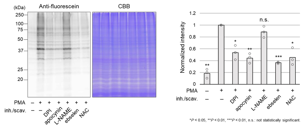
Western blot analysis of the immunostimulation in macrophage cells
RAW264.7 cells were immunostimulated (treated with 1 μg/mL PMA) in the presence of various inhibitors or scavengers described above and incubated for 30 minutes. Afterward, 5 μM Hyp-Stamp was added, and the cells were cultured for 30 minutes. Fluoresein-labeled proteins were assessed by western blot using an anti-fluorescein antibody. The band intensity and the number of labeled proteins increased upon the immunostimulation, and this was decreased by adding inhibitors or scavengers except for L-NAME.
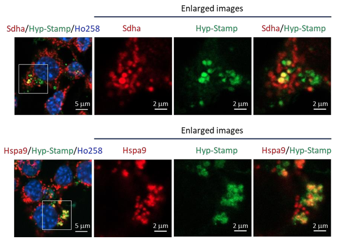
Multistainig of the fluorescein-labeled proteins with antibodies in immunostimulated macrophage cells
RAW264.7 cells were immunostimulated (treated with 1 μg/mL PMA) and incubated for 30 minutes. Afterward, 5 μM Hyp-Stamp was added, and the cells were cultured for 30 minutes. Then, cells were fixed with cold methanol or 4% formaldehyde and immunostained with mitochondrial proteins Sdha and Hspa9. Confocal laser scanning microscope imaging shows the immunofluorescence against Sdha and Hspap partially merged with fluorescein-labeled vesicles.
Imaging of the H2O2 localization in mouse hippocampal slices
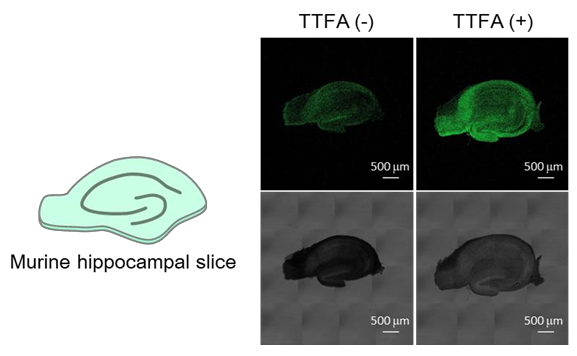
Murine hippocampal slices were treated with mitochondrial Complex II inhibitor TTFA (1 mM, 30 min) to induce oxidative stress, then 5 μM Hyp-Stamp was added and incubated for 1 hour. The slices were fixed with 4% formaldehyde and permeabilized with cold methanol, followed by fluorescent imaging with a confocal laser scanning microscope. Localization of TTFA-triggered H2O2 was observed in the hippocampal slices.
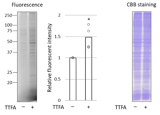
Analysis of total fluorescein-labeled proteins under oxidative stress of mitochondria
HeLa cells were treated with mitochondrial Complex II inhibitor TTFA (1 mM, 30 min) to induce oxidative stress, then 5 μM Hyp-Stamp was added and incubated for 30 min. After preparing a cell lysate, samples are applied to an SDSPAGE, imaged by Coomassie Brilliant Blue (CBB), and ingel fluorescence. Results revealed the amount of fluorescein-labeled protein increased by TTFA treatment, while TTFA did not affect the amount of total protein.
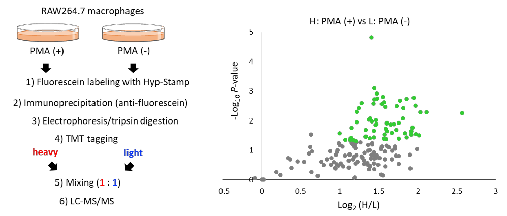
Identification of proteins existing nearby H2O2 generating site by proteomics methodology.
5 μM Hyp-Stamp was added to RAW264.7 cells in which immunostimulation (treated with 1 μg/mL PMA for 30 minutes) was given (+) or not (-), and the cells were cultured for 30 minutes. After preparing the cell lysates, immunoprecipitation was performed with an anti-fluorescein antibody to enrich fluorescein-labeled proteins. Proteins obtained by immunoprecipitation were applied to in-gel trypsin digestion to produce peptide solutions. After modifying the PMA(+) and PMA(-) samples with TMT heavy tag and TMT light tag, respectively, the two samples were mixed in a 1:1 ratio and subjected to LC-MS/MS analysis to investigate changes between samples comprehensively. Figures show the experimental scheme of proteomics analysis (left) and volcano plot of protein signal changes by PMA(+)/(-) (right). In the plot, proteins with log2(PMA added/not added) > 1, and P value < 0.05 are shown in green dots. Mitochondrial proteins such as Sdha and many vesicle proteins were identified, suggesting that these proteins exited nearby H2O2 generated by immunostimulation.
Product Background
Reactive Oxygen Species (ROS) are reactive molecules produced in a wide range of biological processes. ROS are mainly produced during oxidative stress and damage lipids, proteins, and DNAs, which are concerned with cellular senescence and cause various diseases, such as cancer, inflammation, cardiovascular diseases, and neurodegenerative diseases. On the other hand, ROS are also generated in normal metabolic processes and play essential roles in cellular functions by regulating many signaling pathways. Hydrogen peroxide (H2O2) is one of the ROS and exhibits mild reactivity and chemical stability compared with other ROS. Therefore, H2O2 is an ideal cellular signaling molecule that can move and diffuse with sufficient distance. H2O2 regulates various physiological processes, including inflammation reactions and growth factor stimulation. For these reasons, investigating the H2O2 behavior in diverse types of cells has been an important topic in life science.
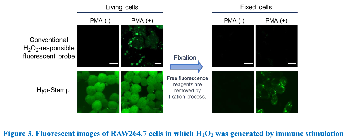
Researchers conventionally use H2O2-responsive fluorescent probes that turns “ON” by reacting with H2O2 to analyze the cellular H2O2. Although they are valuable tools for observing H2O2 generation in living cells, they diffuse to other regions in the cell after reacting with H2O2, which makes it difficult to observe the exact localization of H2O2 generation over time. In addition, the fluorescent probes cannot be used for multistaining with antibodies because they are removed in cell fixation process.
“Hyp-Stamp” is an H2O2-responsive protein labeling reagent. Hyp-Stamp responds to cellular H2O2 and accomplishes fluorescein labeling of proteins lolalized nearby H2O2. Since Hyp-Stamp-mediated fluorescein-labeled proteins remain at the original site, and after cell or tissue fixation, users can observe the H2O2 localization in fixed cells or fixed tissues by fluorescent microscope. Further, performing multistaining of the fluorescein-labeled proteins with antibodies for the protein of user interest is compatible. In addition, proteins localized nearby cellular H2O2 can be analyzed and identified using proteomics methodologies. Hyp-Stamp is a novel and innovative reagent for monitoring the behavior of cellular H2O2 and can be used complementary to conventional fluorescent probes.
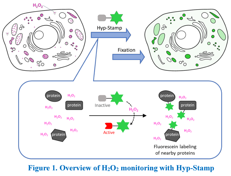
Product principle
“Hyp-Stamp” is a reagent developed by Prof. Hamachi and co-workers at Kyoto University.1 Hyp-Stamp consists of a protein labeling moiety and a Fluorescein moiety. Before reacting with H2O2, the protein labeling moiety is protected with boronic acid ester, which does not react to proteins in this “inactivate form.” The protein labeling portion converts to its “active form” (quinone methide form) by reacting with cellular H2O2, removing the protecting group. The “active form” moiety rapidly reacts with the nucleophilic groups of proteins that exist nearby cellular H2O2.

Conventional H2O2-responsive fluorescent probes can observe H2O2 generation in living cells while they become undetectable in fixed cells because they are removed in the fixation process. On the other hand, Hyp-Stamp can visualize the local site of H2O2 after fixation of the cells, while it cannot observe H2O2 localization in living cells before fixation because of the overwhelmed fluorescence derived from the excess unreacted Hyp-Stamp.
Reference
1. Zhu et al., J. Am. Chem. Soc., 142, 15711–15721 (2020) Imaging and Profiling of Proteins under Oxidative Conditions in Cells and Tissues by Hydrogen-Peroxide-Responsive Labeling.
Related products
- LipiRADICAL Green <Lipid Radical Detection Reagent>
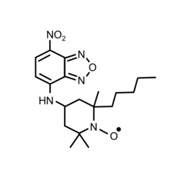
LipiRADICAL Green is a specific fluorescent dye for lipid-derived radicals which are the most upstream factor of lipid peroxidation (LPO). LipiRADICAL Green can be applied into both in vitro assay and cell-based assay to monitor lipid radical productions.
Catalog No. FNK-FDV-0042 .png)
- AcroleinRED <Cell-based Acrolein Detection Reagent>
Acrolein is a LPO downstream aldehyde and one of the most toxic oxidative stress marker. AcroleinRED is the world first cell-based acrolein detection reagent.
Catalog No. FNK-FDV-0022
Catalog No. FNK-FDV-0022
- PolyamineRED <Indracellular Polyamine Detection Reagent>
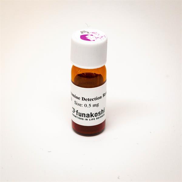
Polyamines are one of the essential class of metabolites in al living organisms and show an anormous number of biological functions. PolyamineRED is the world first reagent for detecting intracellular polyamines without any pre-treatment and cell lysis.
Catalog No. FNK-FDV-0020
Catalog No. FNK-FDV-0020
Related Products
LipiRADICAL Green (Lipid Radical Detection Reagent)
$454.48
$454.48

AcroleinRED (Cell-based Acrolein Detection Reagent)
$454.48
$454.48
PolyamineRED (Intracellular Polyamine Detection Reagent)
$454.48
$454.48
Recently Viewed
Biofactory Accessories
$0.00
MCD Medium
$0.00
WEPRO7240G Core Kit
$3210.27
Cover Glass
$0.00


TN-cyclon ELISA Development Kit
$2095.06
L-Sorbose, ≧99.5%
$153.48