LipiRADICAL Green (Lipid Radical Detection Reagent)
LipiRADICAL Green
Lipid Radical Detection ReagentLipid Peroxidation Innovative Tool
Description
LipiRADICAL Green is the world's first detection reagent for lipid radicals, which are upstream factor of lipid-peroxidation (LPO). This reagent is compatible with live cell imaging, structural analysis of lipid radicals by fluorescent LC/MS-MS and etc.
| Catalog Number | : FNK-FDV-0042 |
| Size | : 0.1 mg |
| Formulation | : C19H28N5O4 |
| Chemical structure | 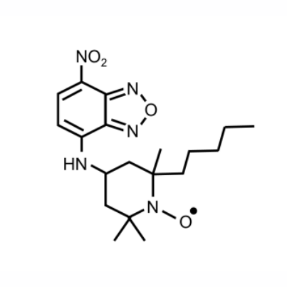 |
| Molecular weight | : 390.21 g/mol |
| Solubility | : Soluble in DMSO |
| Ex/Em | : 470 nm/ 520-600 nm (maximum ~540 nm) |
|
*Conventional FITC filter sets are compatible.
|
|
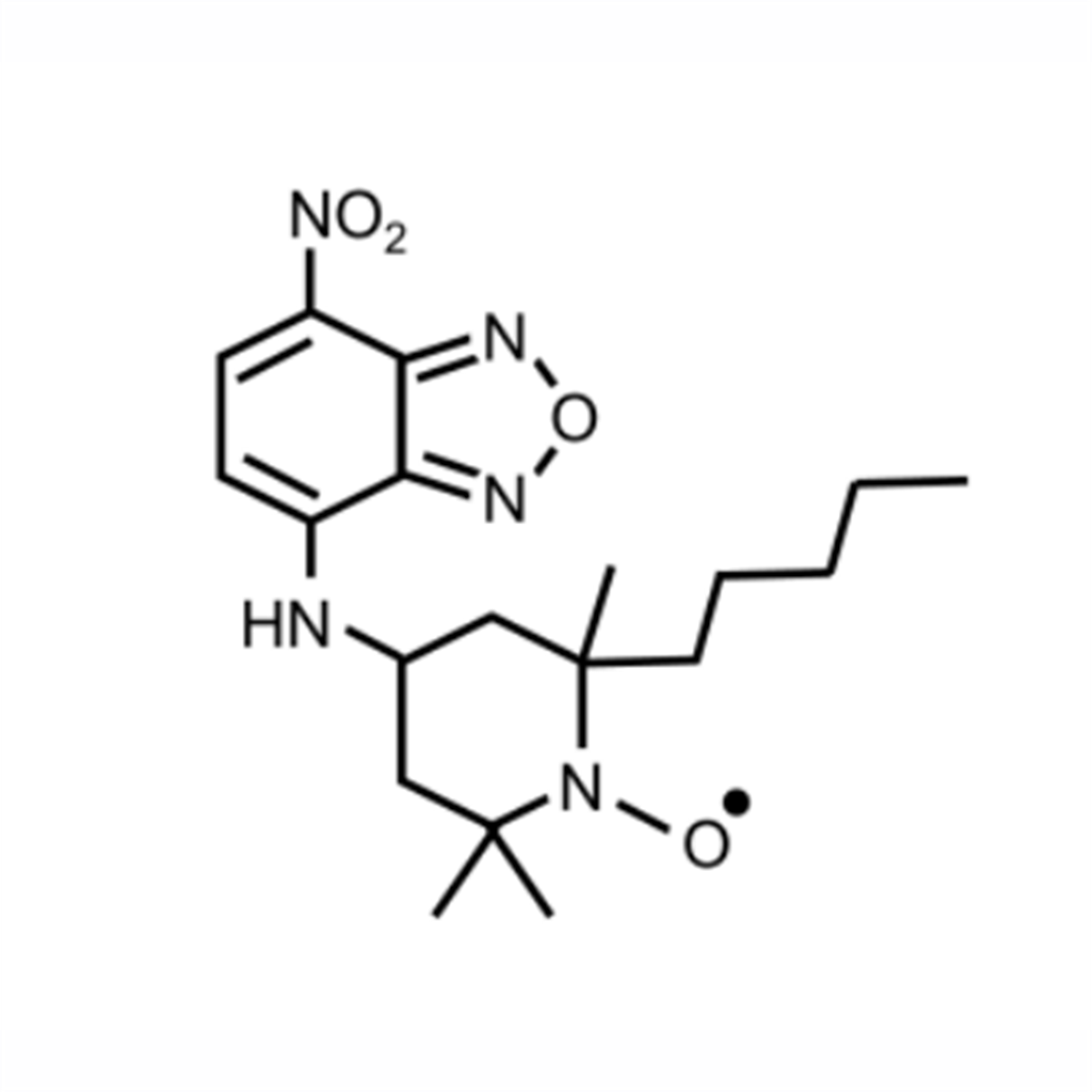
| "LipiRADICAL Green” is the world’s first lipid radical responsive fluorescent dye. “LipiRADICAL Green” is a green fluorescent dye NBD-conjugated nitroxyl radical-derivative (Below figure). Although this compound contains NBD, the probe ’s fluorescence is highly quenched by intramolecular radical moiety. When the probe reacts with lipid radicals via radical-radical coupling forming a covalent bond to lipids, the fluorescent intensity is drastically recovered. “LipiRADICAL Green" is well validated to selectively detect lipid radicals, not reactive oxygen radicals. “LipiRADICAL Green" enables us to semi-quant ification of lipid radicals in biological samples, to image cellular lipid radicals and to identify and analyze the molecular structure of lipid radicals with LC/MS system. “LipiRADICAL Green" is an innovative and powerful tool for LPO research. |
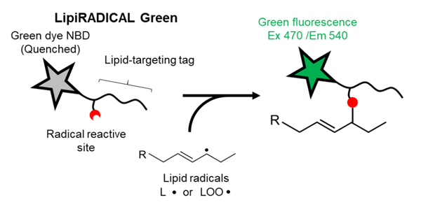
Principal of LipiRADICAL Green
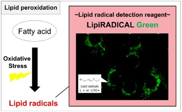
Product Background
Lipid peroxidation (LPO) is one of the several degradation processes of lipids under oxidative stress (Figure 1).Primary products in LPO are lipid radicals and there are two major initiators to induce LPO process, pro-oxidants and lipid oxidative enzymes, including lipoxygenase (LOX) and cytochrome P450 (CYP).
LPO process (1): Pro-oxidants
For pro-oxidantinduced LPO, lipids containing unsaturated fatty acid, especially polyunsaturated fatty acids (PUFAs), are attacked by pro-oxidants, including reactive oxygen species (ROS) and form lipid-derived radicals. Lipid radical (L • ) can be easily oxidized to lipid peroxyl radical (LOO • ). Unstable LOO • immediately extracts a hydrogen from another lipid molecule generating a lipid hydroperoxide (LOOH) and a new lipid radical (L • ).
LPO process (2): Lipid oxidative enzymes
Another pathway enzymeinduced LPO, lipids containing PUFAs are oxidized to lipid hydroperoxides (LOOH), which decomposes to lipid peroxyl radicals LOO • or alkoxyl radicals LO • by metal ions (Fe2+ etc.).
Once lipid radical is produced by the above two processes, lipid radicals expand the radical chain reaction (radical propagation step). In the termination reaction, antioxidants donate a hydrogen atom to the lipid peroxy radical (LOO • ) species resulting in the formation of many different aldehydes, including malondialdehyde (MDA), acrolein, propanal, hexanal, and 4-hydroxynonenal (4-HNE). These aldehydes are cytotoxic because reactive aldehydes attack biomolecules (proteins, DNA/RNA, etc.) to form secondary products. These reactive aldehydes are considered causative factors of organ injury, ferroptosis and ER-stress. To understand the molecular mechanism and physiological relevance of LPO, detection and quantification methods for lipid radicals are required. However, the conventional detection methods are highly limited. For example, electron spin resonance (ESR) is a major strategy to detect radical products but not applied to cell-based applications.
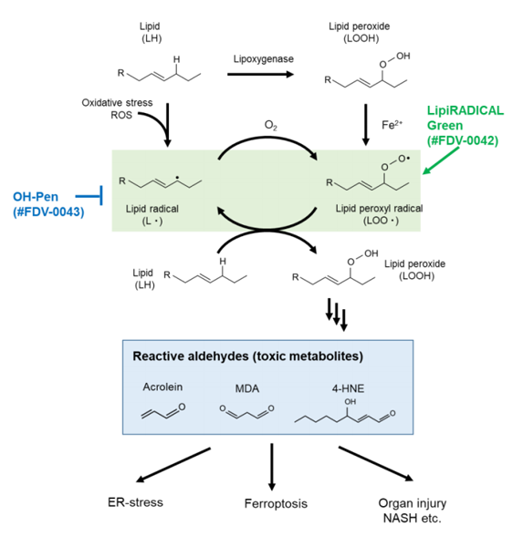
Overview of lipid radicals in LPO pathway
Application
- In vitro detection of lipid radicals by fluorescent detection
- Live cell imaging of lipid radicals by fluorescent microscopy
- Screening of LPO suppressor or antioxidant both in vitro or in cellulo
- Structural analysis of lipid radicals by fluorescent-LC/MS-MS
Reconstitution and Storage
Storage : Store powder at -20°C.
After reconstitution in DMSO, aliquot and store at -20 °C, avoid repeated freeze-thaw cycles. Protect from light.
Reference data
| Fluorescent spectrum | |
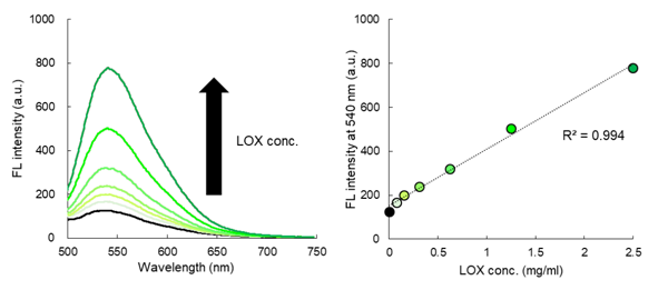
“LipiRADICAL Green” was added to arachidonic acid-lipoxygenase (LOX) mixtures and observed fluorescence excited by 470 nm light. In the absence of LOX enzyme, the fluorescent signal was highly quenched (Black line). In the presence of LOX enzyme, green fluorescence (500-650 nm, maximum ~540 nm) was detected in LOX dosedependent manner |
|
 |
|
| Specificity | |
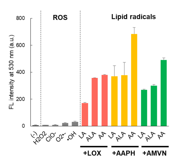
“LipiRADICAL Green” was treated with the following reagents and fluorescent intensity (Ex 470 nm/Em 530 nm) was observed. All reactive oxygen species had little effects on the fluorescent intensity of “LipiRADICAL Green”. Green fluorescence was only observed under the polyunsaturated lipids (laulic acid (LA), α-laulic acid (ALA) or arachidonic acid (AA)) with LOX enzyme or pro-oxidants including AAPH and MeO-AMVN. Reagents and conditions LipiRADICAL Green (5 μM) H2O2, ClO-, KO2 for O2 - • and • OH : 0.5 mM Lipids (0.5 mM) with LOX (2.5 μg/ml) , AAPH (10 mM) or MeO-AMVN 50 μM
|
|
 |
Application data
| Cell-based detection of lipid radicals induced by diethylnitrosamine (DEN) | |
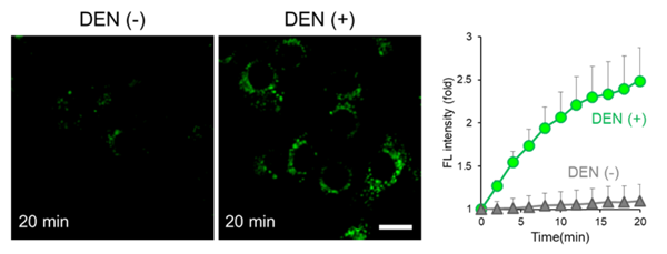
Hepa1-6 cells were treated with 1 μM of “LipiRADICAL Green” for 20 min and washed twice with PBS. For inducing an LPO signal, the cells were co-treated with diethylnitrosamine (DEN) and “LipiRADICAL Green”, an LPO initiator. Immediately after DEN addition, the cells were observed by confocal microscopy (Ex.458 nm/ Em. 490-674 nm) for 20 min with 2 min interval. The fluorescent signal of “LipiRADICAL Green” from the DENtreated cells clearly increased. |
|
 |
|
| in vitro detection of lipid radicals derived from LDL | |
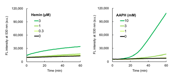
Purified low-density lipoprotein (LDL, 20 μg protein/mL) was mixed with pro-oxidants hemin or AAPH and “LipiRADICAL Green” and the green fluorescence (Ex. 470 nm/ Em 530 nm) was measured for 60 min at 37oC. Both hemin and AAPH increased green fluorescence indicating the production of lipid radicals from LDL particles in a time-dependent manner. |
|
 |
|
| Structural analysis of lipid radicals derived from arachidonic acid in vitro | |
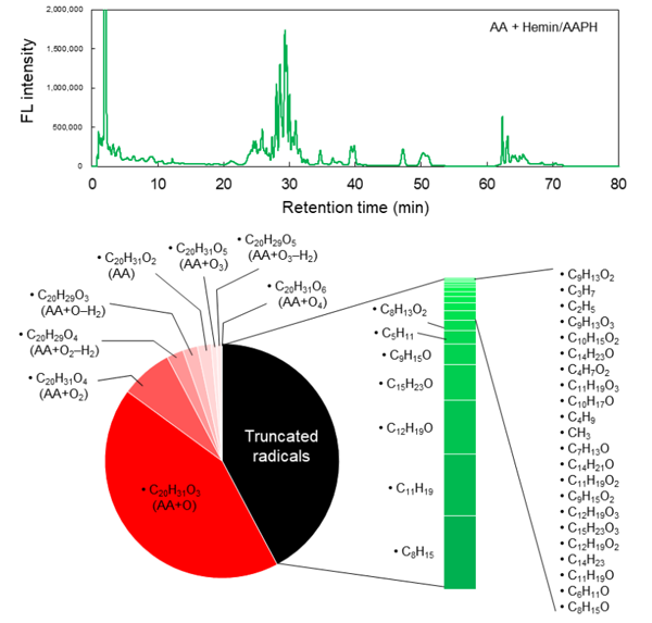
Arachidonic acid (AA; 500 μM) was incubated with pro-oxidants hemin (10 μM) and AAPH (50 mM) mixture for 60 min. After incubation, 5 μM of “LipiRADICAL Green” was added to the reaction mixture and incubated for 15 min at R.T. Lipid components were extracted by the Bligh and Dyer method and analyzed by the LC-FL/MSMS technique. (Upper panel) The fluorescent chromatogram is shown (Ex. 470/E. 530 nm). Several fluorescent peaks were observed and each peak was further analyzed by MS-MS. (Lower panel) Product profiles of AAderived radicals are shown. MS-MS analysis identified a total of 8 full-length AA radicals and 29 truncated radicals. The relative abundances of each radical were calculated from each peak area. Detailed experimental protocol and analytical procedure are described in Ref.5. |
|
 |
|
| Structural analysis of lipid radicals in vivo | |
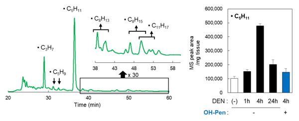
A well-known carcinogen, diethylnitrosamine (DEN, 100 mg/kg body weight), was injected intraperitoneally into mice and after 1, 4 and 24 hours, mice were anesthetized. Anesthetized mice then received intraperitoneal injections of “LipiRADICAL Green” (2.5 μmol/kg body weight). To check the specificity of “LipiRADICAL Green”, OHPen, a specific inhibitor of lipid radical (Catalog no. #FDV-0042; 10 μmol/kg body weight) was also injected into the mice before “LipiRADICAL Green” injection. The liver was removed from the mice and homogenized with methanol. Lipid solution was extracted from the liver homogenate according to the Bligh and Dyer method. Lipid samples were applied to LC-FL/MS-MS for analysis (Left). After 4 hours of treatment of DEN, there was a high production of lipid radicals. A total of 11 lipid radicals were identified. (Right) An example, a • C5H11 radical. OH-Pen-preinjection clearly inhibited the production of lipid radicals derived from DEN treatment. |
|
 |
Based on the technical data and publication history provided, LipiRADICAL Green is utilized by researchers primarily as a highly specific tool for detecting lipid radicals (the initiators of lipid peroxidation) in various biological contexts, ranging from molecular analysis to in vivo imaging.
Here is a summary of how researchers utilized the reagent across the cited publications:
1. Visualization & Live-Cell Imaging
A primary use of LipiRADICAL Green is to visualize the spatial and temporal production of lipid radicals in live cells.
-
Ferroptosis Studies: Researchers (e.g., Mishima et al. 2022, Lange et al. 2025) used the probe to monitor the onset of ferroptotic cell death, specifically identifying where lipid radicals accumulate before membrane rupture.
-
Organelle-Specific Stress: Studies like Shiiba et al. (2025) utilized it to track radical transfer between the ER and mitochondria, while Miki et al. (2024) focused on lysosomal dysfunction and lipid radical buildup during glucose starvation.
-
Cancer Research: In studies regarding Triple-Negative Breast Cancer and Glioblastoma (Mathew et al. 2024, Miki et al. 2022), it was used to demonstrate how specific drugs (like Niclosamide) or metabolic conditions trigger lethal oxidative stress.
2. Structural Analysis (LC-FL/MS-MS)
Unlike general oxidative stress markers, LipiRADICAL Green allows for the identification of the exact molecular species of the radical.
-
Radical Fingerprinting: Matsuoka et al. (2020) and Yamada et al. (2016) utilized the probe's ability to form stable covalent bonds with lipid radicals. By extracting these complexes and analyzing them via LC/MS-MS, researchers identified the specific carbon-centered radicals (e.g.,C?H₁₁ radicals) formed from polyunsaturated fatty acids like arachidonic acid.
3. In Vivo and Tissue-Level Detection
The probe has been successfully applied to animal models to study systemic disease processes.
-
Retinal & Cardiac Damage: Enoki et al. (2017) used it to prove that lipid radicals are the direct cause of light-induced retinal degeneration. Similarly, Yagi et al. (2023) and Do et al. (2024) used it to detect radical production in the heart to study heart failure and mitochondrial transporter deletions.
-
Systemic Carcinogens: Researchers used the probe in mice to image the liver following Diethylnitrosamine (DEN) injection, showing real-time production of lipid radicals in situ.
4. Screening for Antioxidants & Suppressors
LipiRADICAL Green serves as a benchmark for testing the efficacy of new drugs or internal cellular defense mechanisms.
-
Antiferroptotic Agents: Mishima et al. (2020) used the probe to screen for repurposed drugs that function as radical scavengers to suppress acute kidney injury (AKI).
- Metabolic Defense: Shinjyo et al. (2025) used it to evaluate how aldehyde metabolism protects lung mucociliary clearance from air pollution-induced oxidative damage.

References
- Yamada et al., Nat. Chem. Biol., 12, 608-613 (2016) Fluorescence probes to detect lipid-derived radicals.
- Enoki et al., Chem. Commun., 53, 10922-10925 (2017) Lipid radicals cause light-induced retinal degeneration.
- Ishida et al., Free Radical Biol. Med., 113, 487-493 (2017) Detection and inhibition of lipid-derived radicals in low-density lipoprotein.
- Mishima et al., J. Am. Soc. Nephrol., 31, 280-296 (2020) Drug Repurposed as antiferroptosis agents suppress organ damage, including AKI, by functioning as lipid peroxyl radical scavengers.
- Matsuoka et al., Anal. Chem., 92, 6993-7002, (2020) Method for structural determination of lipid-derived radicals
- Mishima, E., et al., Nature, 608 (7924), 778~783 (2022) A non-canonical vitamin K cycle is a potent ferroptosis suppressor.
- Mathew, M., et al., Antioxidants (Basel). 13(3):291 (2024) Induction of Oxidative Stress and Ferroptosis in Triple-Negative Breast Cancer Cells by Niclosamide via Blockade of the Function and Expression of SLC38A5 and SLC7A11
- Lange, M., et al. (2025). FSP1-mediated lipid droplet quality control prevents neutral lipid peroxidation and ferroptosis. Nature Cell Biology, 27(11), 1902–1913.
- Miki, K., Yagi, M., Yoshimoto, K., Kang, D., & Uchiumi, T. (2022). Mitochondrial dysfunction and impaired growth of glioblastoma cell lines caused by antimicrobial agents inducing ferroptosis under glucose starvation. Oncogenesis, 11(1), 59. https://doi.org/10.1038/s41389-022-00437-z
- Shiiba, I., Ito, N., Oshio, H., et al. (2025). ER-mitochondria contacts mediate lipid radical transfer via RMDN3/PTPIP51 phosphorylation to reduce mitochondrial oxidative stress. Nature Communications, 16, 1508. https://doi.org/10.1038/s41467-025-56666-4
- Miki, K., Yagi, M., Kang, D., Kunisaki, Y., Yoshimoto, K., & Uchiumi, T. (2024). Glucose starvation causes ferroptosis-mediated lysosomal dysfunction. iScience, 27(5), 109735. https://doi.org/10.1016/j.isci.2024.109735
- Yagi, M., Do, Y., Hirai, H., Miki, K., Toshima, T., Fukahori, Y., Setoyama, D., Abe, C., Nabeshima, Y. I., Kang, D., & Uchiumi, T. (2023). Improving lysosomal ferroptosis with NMN administration protects against heart failure. Life Science Alliance, 6(12), e202302116. https://doi.org/10.26508/lsa.202302116
- Do, Y., Yagi, M., Hirai, H., Miki, K., Fukahori, Y., Setoyama, D., Yamamoto, M., Furukawa, T., Kunisaki, Y., Kang, D., & Uchiumi, T. (2024). Cardiomyocyte-specific deletion of the mitochondrial transporter Abcb10 causes cardiac dysfunction via lysosomal-mediated ferroptosis. Bioscience Reports, 44(5), BSR20231992. https://doi.org/10.1042/BSR20231992
- Loo, T. M., Zhou, X., Tanaka, Y., Sugawara, S., Yamauchi, S., Kawasaki, H., ... & Takahashi, A. (2025). Senescence-associated lysosomal dysfunction impairs cystine deprivation-induced lipid peroxidation and ferroptosis. Nature Communications, 16, 6617. https://doi.org/10.1038/s41467-025-61894-9
- Unten, Y., Takafuji, K., Masukagami, Y., Shiiba, I., Horiuchi, K., Husnik, F., Yanagi, S., Tateishi, N., & Suzuki, T. (2026). Characterizing mitochondrial phenotypes and MERCS in aged human skeletal muscle myoblasts. PLOS One, 21(2), e0343604. https://doi.org/10.1371/journal.pone.0343604
- Hirata, Y., Yamada, Y., Taguchi, S., Kojima, R., Masumoto, H., Kimura, S., ... & Matsuzawa, A. (2024). Conjugated fatty acids drive ferroptosis through chaperone-mediated autophagic degradation of GPX4 by targeting mitochondria. Cell Death & Disease, 15(12), 884. https://doi.org/10.1038/s41419-024-07237-w
- Shinjyo, N., Kimura, H., Yoshihara, T., Suzuki, J., Yamaguchi, M., Kawabata, S., & Okabe, Y. (2025). Aldehyde metabolism governs resilience of mucociliary clearance to air pollution exposure. The Journal of Clinical Investigation, 135(14), e191276. https://doi.org/10.1172/JCI191276
- Nguyen, N. T., Sennoune, S. R., Dharmalingam-Nandagopal, G., Sivaprakasam, S., Bhutia, Y. D., & Ganapathy, V. (2024). Impact of Oncogenic Changes in p53 and KRAS on Macropinocytosis and Ferroptosis in Colon Cancer Cells and Anticancer Efficacy of Niclosamide with Differential Effects on These Two Processes. Cells, 13(11), 951. https://doi.org/10.3390/cells13110951