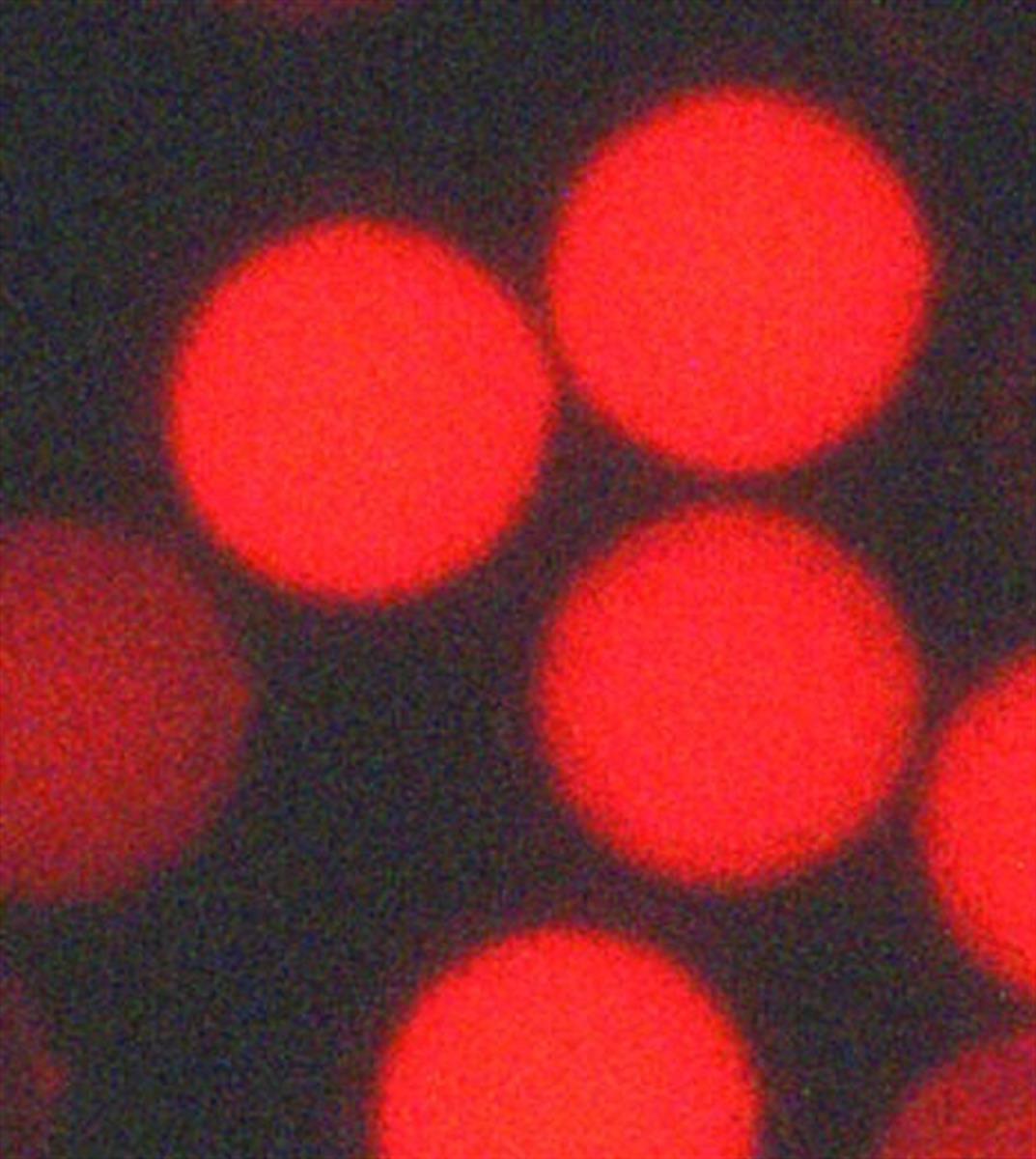
No More Sales-Flamma® Deep Red PS Bead 0.2 um
Packing Unit
-
2 mL

-
5 mL

-
10 mL

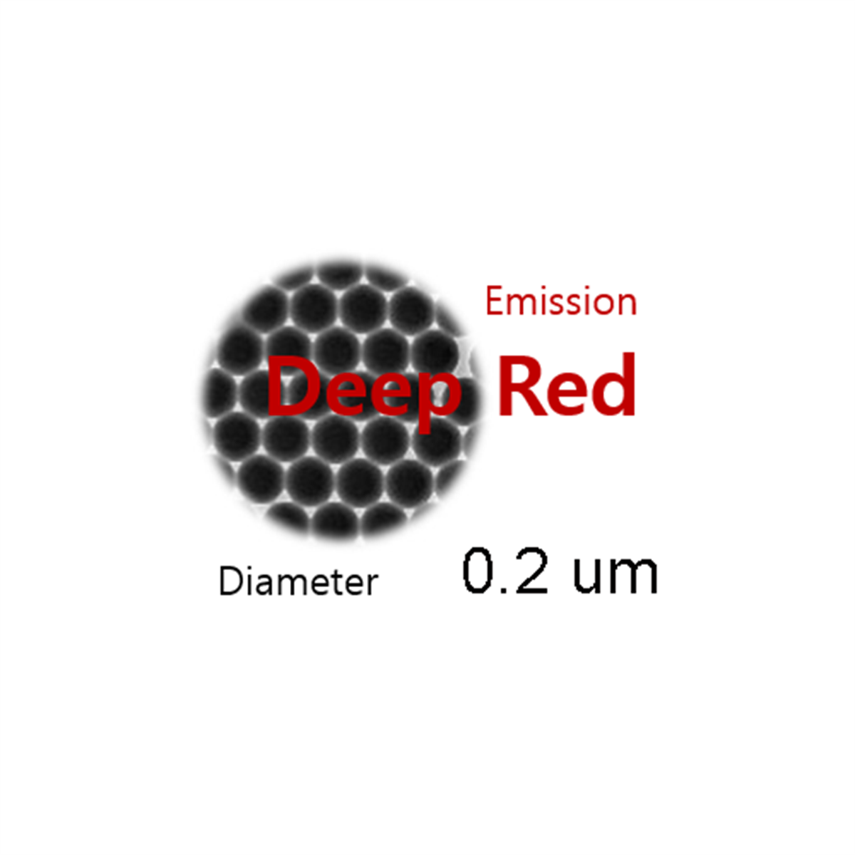
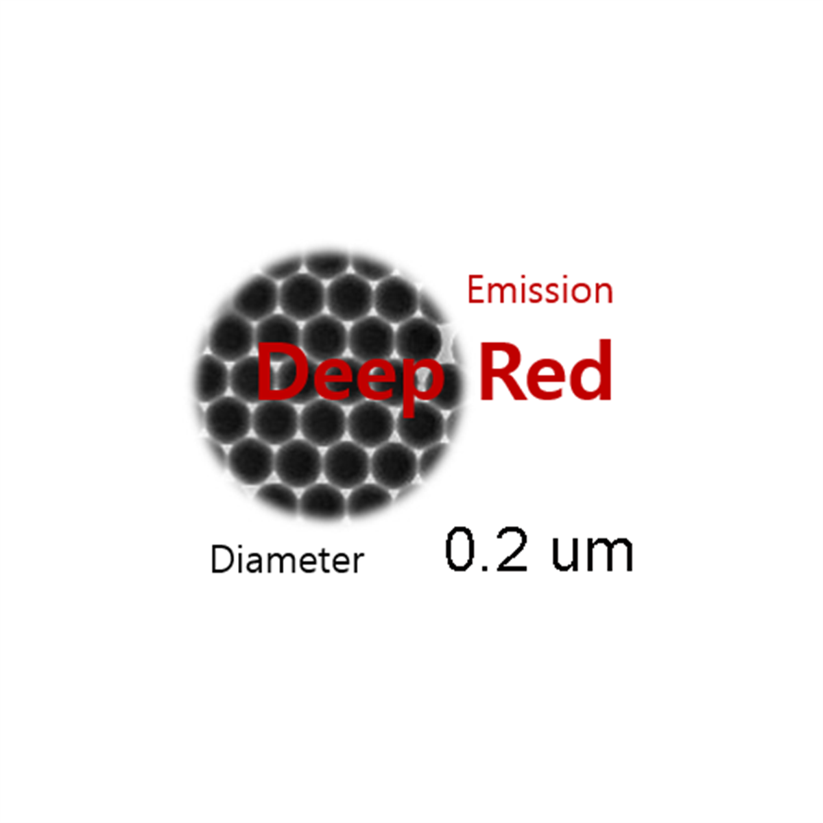
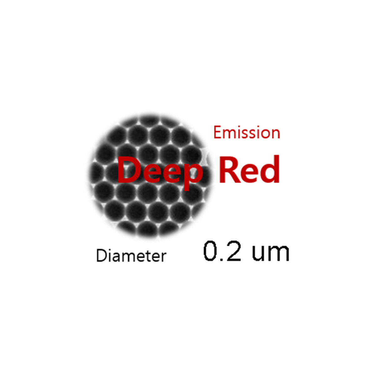
Flamma® Deep Red PS Bead 0.2 um
Cat. No. List below
Description
The carboxylic acid-modified surface allows for covalent coupling with proteins and amine-containing biomolecules, easy functionalization using water-soluble coupling reagents, and customizable surface properties for specific applications.
Flamma® Deep Red PS Beads are suitable for nanoparticle-based diagnostic tests, particle and cell tracking, fluid dynamics tracin, In vivo uptake and transport studies, flow cytometry, and fluorescence microscopy.
It has deep red fluorescence minimizes autofluorescence interference, has high signal-to-noise ratio for sensitive detection, and excellent photostability for extended imaging sessions. It has uniform size and shape by having a precise 0.2 μm diameter for consistent results and its spherical morphology makes it ideal for fluid dynamics studies and cellular uptake experiments.
Flamma® Deep Red PS Bead 0.2μm represents a significant advancement in fluorescent nanoparticle technology, offering researchers a versatile and high-performance tool for a wide range of applications in biomedical research, diagnostics, and materials science. Their unique combination of size, surface chemistry, and optical properties makes them ideal for both in vitro and in vivo studies, pushing the boundaries of what's possible in nanoscale imaging and detection.
Unique advantages:
1. Proprietary Dye Technology: Flamma® series dyes offer enhanced brightness and stability compared to conventional fluorophores
2. Size-Dependent Studies: The 0.2 μm diameter is optimal for investigating size-dependent phenomena in biological systems
3. Biocompatibility: High-grade polystyrene core ensures low toxicity and minimal interference with biological processes
4. Versatile Excitation: Compatible with common laser lines and LED light sources in the red spectrum
5. Stable Suspension: 2% solids content provides a good balance between particle concentration and suspension stability
Specifications
- Fluorophore: Flamma® Deep Red
- Surface modification: Carboxylic acid
- Excitation/Emission Max.(nm): 645/665 nm
- Appearance: Aqueous suspension
- Bead diameter: 0.2 μm
- Solids: 2% (m/v)
- Storage conditions: 4℃, protect from light
Table 1. Flamma® PS Bead series list
| Quick link (Cat.#) | Series | Ex * (nm) | Em* (nm) | Bead size | Functionality | Solids |
| PSC7003 | Flamma® Deep Red PS Bead 0.1 um | 645 | 665 | 0.1 μm | COOH | 2% |
| PSC7001 | Flamma® Deep Red PS Bead 0.2 um | 645 | 665 | 0.2 μm | COOH | 2% |
| PSC7002 | Flamma® Deep Red PS Bead 0.5 um | 645 | 665 | 0.5 μm | COOH | 2% |
Overview
Flamma® PS bead series are fluorescent spherical particles in the colloidal size made up of high-grade polystyrene (PS) doped with our proprietary dyes. Polystyrene beads are not cytotoxic, and they diffuse minimally from the injection site and persist for long periods in nerve cells. Our fluorescent beads display excellent optical properties yet negligible photobleaching under the intense excitation for fluorescence microscopy. The intense fluorescence and spherical shape of Flamma® PS bead series enable to generate stronger signals with small amount of probes in many tracing experiments. To decrease nonspecific binding and provide additional functional groups for conjugation, exteriors of PS bead are equipped with carboxylic acids. These pendent carboxylic acids allow them suitable for covalent coupling of proteins and other aminecontaining biomolecules by using water-soluble coupling reagents. BioActs provides various sizes of Flamma® PS bead series (0.1 – 0.5 μm) in order to meet the diverse needs of our customer, and sizes specified in the product names are nominal bead diameters. These effective fluorescent PS beads can be utilized in a variety of applications such as neuronal tracers, cellular antigen markers, cell tracers, and the standardization reagents for flow cytometry. Due to their high fluorescence intensity, Flamma® PS bead series are suitable for the many nanoparticle based diagnostic tests, including tracking particles and cells, tracing fluid dynamics, discriminating the size dependence of uptake or transport in vivo. Although limited sizes of our fluorescent beads are available, we can prepare custom-sized particles, custom-dyed or custom-surface modifications orders upon request.
All Flamma® Fluors PS beads are stable under sonication, vortexing, or shaking process. All Flamma® Fluors PS beads should be stored at 2–6°C, protected from light, and do not freeze. Our Flamma® Fluors PS beads are supplied as suspensions (2% solids) in water containing 0.05% sodium azide. The PS beads are stable for at least one year, provided recommended storage conditions are strictly observed. Before sampling, mix well by sonication, vigorous shaking, or vortex mixing.
Bead activation and amine conjugation
amines of biomolecules. Primary amines such as lysine can react with NHS ester under the physiological pH in aqueous condition.
Protocols for NHS ester activation
2. Add N-hydroxysuccinimide and EDCI into the diluted bead solution, and mixed the mixture for 30 min at rt.
3. Centrifuge the mixture at 15,000 Xg for10 min, and then remove supernatant.
4. Suspend the bead pellet in 50 mM MES buffer (pH 6.4), and repeat the step 3 twice
Protocols for antibody (Goat-anti-mouse IgG) conjugation
2. Add antibody solution to bead suspension solution, and stand the mixture for 4 h at rt
(bead 1 mg per antibody 100 μg)
3. Centrifuge the mixture and remove supernatant, and wash bead pellet three times with 0.01% Tween-20 in PBS buffer
Blocking/Quenching
1x BSA or 1 mM amine solution in PBS after step 2 in conjugation protocol and perform step 3.
Citation & Reference
1. Stability of water-soluble carbodiimides in aqueous solution. Gilles, M.A., Hudson, A.Q., Borders, C.L., 1990. Anal. Biochem. 184, 244-248
2. Different EDC/NHS activation mechanisms between PAA and PMAA brushes and the following amidation reactions. Wang, C., Yan, Liu, H.B., Zhou, X.H., Xiao, S.J., 2011. Langmuir 27 (19), 12058-12068.
3. Mechanism of amid formation by carbodiimides for bioconjugation in aqueous media. Nakajima, N., Ikada, Y., 1995. Bioconjug. Chem. 6, 123-130
4. The adsorptive characteristics of proteins for polystyrene and their significance in solid-phase immunoassays. Cantarero, L.A., Butler, J.E., Osborne, J.W., 1980. Anal. Biochem. 105, 375-382
5. The physical and functional behavior of capture antibodies adsorbed on polystyrene. Butler, J.E., Ni, L., Nessler, R., Joshi, K.S., Suter, M., Rosenberg, B., et al., 1992. J. Immunol. Methods 150, 77-90.
6. N-hydroxysulfosuccinimide active esters: Bis(N-hydroxysulfosuccinimide)ester of two dicarboxylic acids are hydrophilic, membrane impermeant, protein cross-linkers. Staros, J.V., 1982. Biochemistry 21, 3950-3955