PVP Protein Extraction Buffer
PVP Protein Extraction Buffer
A mild, Tris-based protein extraction system with PVP-40 for polyphenol binding and TCEP-HCl for stable protein reduction. Supplied with a separate 100X protease inhibitor cocktail for flexible, plant-optimized protein isolation.
- PVP-fortified formulation (1% PVP-40) to bind polyphenols and reduce pigment and tannin interference during protein extraction from challenging tissues.
- TCEP-HCl provides a stable, thiol-free reducing environment that protects cysteine residues and minimizes oxidative protein damage.
- Tris and NaCl offer a physiological-like environment that maintains protein solubility and native structure for many targets.
- Compatible with common downstream analyses including SDS-PAGE, western blotting, enzyme assays, and total protein quantification.
- Ultrapure Type 1 water (18.2 MΩ·cm)
- Customizable formulation available upon request
- pH7.5 for solution A
- AppearanceClear Solution
- Filtration0.1µm
- Shelf Life1 year
Protein Storage Buffer (Base)
- Storage2-8 °C, avoid repeated freeze-thaw cycles
- Shelf Life1 year
- Tris base50 mM
- Sodium Chloride150 mM
- PVP-401%
- TCEP-HCl5 mM
Protease Inhibitor Cocktail (100X) for Solution A
- Storage-80 °C (long term), avoid repeated freeze-thaw cycles
- Shelf Life1 year
- AEBSF·HCl10 mM
- Aprotinin80 μM
- Bestatin5 mM
- E-64100 μM
- Leupeptin1 mM
- Pepstatin A0.1 mM
Standard laboratory reagents filtered at 0.22 µm leave sub-micron particulates, mycoplasma, and aggregates that interfere with sensitive downstream applications. PVP Protein Extraction Buffer is manufactured under ISO 13485 quality systems with multi-stage ultra-filtration to eliminate these failure modes.
PVP-40 phenol protection
1% PVP-40 efficiently complexes polyphenols and pigments that cause protein crosslinking and assay interference in plant tissues.
Stable TCEP-HCl reduction
5 mM TCEP-HCl maintains cysteine residues in a reduced state and limits oxidative damage during extraction.
Physiological Tris/NaCl base
50 mM Tris and 150 mM NaCl support protein solubility and native structure for many targets.
Broad downstream compatibility
Compatible with SDS-PAGE, Western blotting, enzyme assays, and total protein quantification.
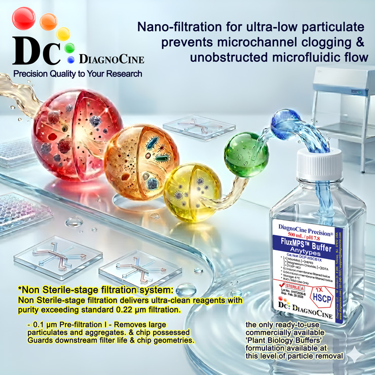
Sequential filtration system
A sequential filtration process reaching 0.1 µm for clean reagent purity exceeding standard 0.22 µm filtration.
-
1
0.1 µm filtration
Removes large particulates, aggregates, and bacteria. Primary guard providing baseline purity exceeding standard 0.22 µm filtration.
Performance vs. conventional reagents
Standard 0.22 µm single-pass filtration leaves particles in the 0.12–0.22 µm range. FluxMPS™ removes these contaminants for cleaner results and higher reproducibility.
Validated applications
- Plant tissues rich in polysaccharides and polyphenols (leaves, roots, seeds, woody tissues).
- Other complex samples where standard guanidinium reagents may give low yield or contaminated RNA (user validation required).
Protocol & usage instructions
- Equilibrate the buffer and samples on ice.
- Immediately before use, thaw and add protease inhibitor cocktail to the buffer.
- Homogenize tissue or cell pellets in 5–10 volumes of cold PVP Protein Extraction Buffer (for example, 0.5-1 mL per 50-100 mg tissue), keeping samples on ice.
- Centrifuge at high speed (e.g., 10,000-20,000 × g, 10-20 minutes, 4 °C) to remove debris.
- Collect the supernatant as the clarified protein extract and use directly for downstream applications or store aliquots at −20 °C or −80 °C as appropriate.
Quality assurance & logistics
ISO 13485:2016 Certified
Manufactured under certified QMS with full traceability and lot documentation.
Made in USA
All manufacturing, QA testing, and final assembly at DiagnoCine Precision, Totowa, NJ.
Supporting literature
- Wang W, Vignani R, Scali M, Cresti M. A universal and rapid protocol for protein extraction from recalcitrant plant tissues for proteomic analysis. Electrophoresis. 2006;27(13):2782-2786.
- Saravanan RS, Rose JKC. A critical evaluation of sample extraction techniques for enhanced proteomic analysis of recalcitrant plant tissues. Proteomics. 2004;4(9):2522-2532.