

MitoFlamma® Green (live)
Product#: RMS1101
$219.36
Packing Unit
-
100Test

Availability:
Ships in 1-2 Weeks
MitoFlamma® Green (live)
Cat. No. : RMS1101_20 X 50 UL | RMS1101_1 ML
Description
MitoFlamma® Green is a mitochondria-selective green fluorescent dye that allows to detect mitochondrial morphology in living cells. This unique dye appears to preferentially accumulate in mitochondria. MitoFlamma® green enables researchers to observe mitochondrial activity, localization and abundance as well as monitoring the effect of drugs or other external stimuli on the mitochondrial function. The maxima of Ex/Em values are at 498/583 nm. We offer MitoFlamma® green as an effective mitochondria specific fluorescent probe for various experiments such as immunocytochemistry, in situ hybridization, microplate-based analysis, etc.
Specifications
- Fluorophore: MitoFlamma® Green
- Application: Cell structure imaging
- Labeling to: Mitochondria
- Excitation/Emission Max.(nm): 498/583
- Molecular weight: 635 g/mol
- Appearance: Red Solution
- Concentration: 1mM
- Solvent: 1% DMSO in PBS or media
- Storage conditions: -20 ℃, protect from light
Background
MitoFlamma® Green is a mitochondria-selective green fluorescent dye that allows to detect mitochondrial morphology in living cells. MitoFlamma® Green appears to preferentially accumulate in mitochondria regardless of mitochondrial membrane potential in certain cell types, making it a possible tool for determining mitochondrial mass. MitoFlamma® Green enables researchers to observe mitochondrial activity, localization and abundance as well as monitoring the effect of drugs or other external stimuli on the mitochondrial function. MitoFlamma® Green probes are retained in the mitochondria during the fixation step and after permeabilization with some detergents during subsequent processing steps. After fixation, labeled samples can be applied in a variety of experiments such as immunocytochemistry, in situ hybridization, microplate-based analysis, etc.
MitoFlamma® Green is a mitochondria-selective green fluorescent dye that allows to detect mitochondrial morphology in living cells. MitoFlamma® Green appears to preferentially accumulate in mitochondria regardless of mitochondrial membrane potential in certain cell types, making it a possible tool for determining mitochondrial mass. MitoFlamma® Green enables researchers to observe mitochondrial activity, localization and abundance as well as monitoring the effect of drugs or other external stimuli on the mitochondrial function. MitoFlamma® Green probes are retained in the mitochondria during the fixation step and after permeabilization with some detergents during subsequent processing steps. After fixation, labeled samples can be applied in a variety of experiments such as immunocytochemistry, in situ hybridization, microplate-based analysis, etc.
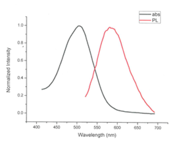
Figure 1. Excitation/emission spectra of MitoFlamma® Green
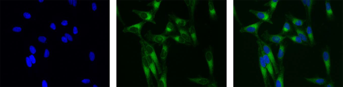
Figure 2. HeLa cell staining with MitoFlamma® Green
Left: DAPI, middle: MitoFlamma Green, right: merged image
Before begining of mitochondrial labeling experiment
Materials and equipment required but not provided
- DMSO
- PBS buffer or suitable growth medium for live cell imaging
- Aldehyde based fixatives such as paraformaldehyde for cell fixation
- Aldehyde based detergents such as Trition® X-100
- Micropipette
- Fluorescence microscope
- 37 ℃ incubator
Labeling of mitochondria in fixed or suspended cells with MitoFlamma® Green
- Prepare the fixed/suspended cells by seeding appropriate cells onto confocal dishes 6 hours before.
- Adjust the temperature of the media to 37 ℃.
- Prepare a 1 μM labeling MitoFlamma® Green solution by diluting original 1 mM DMSO solution with the media from step 3, *protect from light.
- Aspirate the culture medium from the cells grown dishes (for suspended cells, centrifuge the dishes in advance), and add 1 μM labeling MitoFlamma® Green solution to the dishes and incubate for 1 h at 37 ℃ under 5% CO2 in humid condition. *the incubation may be varied for optimal labeling condition.
- Aspirate the labeling media, and rinse the cells twice with PBS at 37 ℃.
- Analyze stained cells with appropriate equipment
Fixation (continuing from step 5 in labeling experiment)
- Fix the cells for 15 min in 4%parnformaldehyde at 37 ℃.
- Aspirate the fixative, and rinse the cell twice with PBS.
- View the cells using FITC filter set.
- When permeabilization is needed for subsequent steps such as immunocytochemistry, incubate fixed cells in buffer containing detergent such as 0.2% Triton® X-100.
- Following permeabilization, rinse the cells in buffer and proceed with immunocytochemistry procedure.
- Alternatively, the cells may be permeabilized by incubating in ice-cold acetone for 5 minutes, and then washed in PBS.
- Even when cells are not going to be labeled with an antibody, this acetone-permeabilization step may be useful in improving the signal-to background ratio.

Recently Viewed

Tray for Pre-Made Agarose Gel
$103.00





Flamma® 552 PEG4-Alkyne
$1632.80
Goat anti-rabbit IgG, FSD™ 647
$339.20
Beauvericin, 1 mg
$87.72