


Imaging Probe Types
Product#: DC-Imaging-Probe-Types
$0.00
Select BIOACT
- Check below icons
Availability:
Ships in 24 hours
Some of items are currently out of stock. Please submit an inquiry if you would like to check availability or make a purchase
Inquire about the Item to, support@diagnocine.comIn Vivo Imaging Probes (Fluorescence)
NpFlamma® HGC series NpFlamma® HGC series is a fluorescent dye incorporated biocompatible and biodegradable chitosan based amphiphilic nanoparticle that enables to selectively detect tumor cells. Chitosan nanoparticles can selectively accumulate in cancer tissues due to high permeability for loose new blood vessels around cancer tissues and retention effect. The polymeric nanoparticles form self-aggregates size of several hundred nanometers in the aqueous system accumulate only in vicinity of cancer tissues. Chitosan particles display low toxicity along with absence of noticeable side effect in vivo, yet they exhibit a long half-life, high stability and aqueous solubility. Thus, NpFlamma® HGC series is an ideal fluorescence agent for in vivo imaging of angiography and tumor progression. In conjunction with NIR dyes, the series allows to observe non-invasive images of cancer metastases and to contrast blood vessel. The hydrophobic nature of NpFlamma® HGC series can embed hydrophobic materials, thus they can be utilized as a selective carrier for hydrophobic cancer drugs such as doxorubicin and paclitaxel, etc. In addition, carboxylic acids on the surface of NpFlamma® HGC series can bind to a variety of biomolecules via amide or ester linkages enabling to utilize them in multi-purposes. NpFlamma® MMP series Matrix metalloproteinases (MMPs) are a group of calcium-dependent, zinc-containing endopeptidases that that in concert are responsible for the degradation of most extracellular matrix (ECM) proteins during organogenesis, growth and normal tissue turnover. Humans have 23 MMPs family, and the expression and activity of MMPs in adult tissues is normally quite low, but increases significantly in various pathological conditions that may lead into unwanted tissue destruction, tumor growth and metastasis, inflammatory diseases, etc. Cancer cells secrete VEGF and FGF in order to induce angiogenesis in vascular endothelial cells, but cannot reach to vascular endothelial cells as being caught in ECM. Although MMPs are connected with cancer cells survival and expansion, only very small amount of MMPs are synthetized by cancer cell. However, the cancer cells induce inflammatory cells to secrete MMPs for assisting the delivery of VEGF secreted by cancer cells and make peripheral space for angiogenesis. The connection between MMPs and apoptosis, cell migration and angiogenesis enables to utilizing MMPs as tumor markers. An increased expression and activity of MMPs in both tissues and blood of patients with various types of cancer is observed. Annexin V Flamma® series Annexin V Flamma® series is a rapid and selective probe for labeling of externalized PS, an indicator of intermediate stages of apoptosis. Flamma® dyes display strong absorption, high fluorescence quantum yield and high photostability, and they maintain good fluorescence activity and stability after conjugation to biomolecules, allowing the detection of low-abundance biological structures with great sensitivity. Annexin V Flamma® conjugates can be utilized in fluorescent imaging as well as for flow cytometry of apoptotic cells, and they might be used in combination with other dyes such as propidium iodide (PI) in order to accurately analyze mixed populations of apoptotic and other cells. Annexin V Flamma® probes available as stand-alone reagent or easy-to-us kits. ApoFlamma® series ApoFlamma® H series made up of a covalent-connection of CQRPPR hexapeptide (ApoPep) with corresponding fluorophore. ApoPep strongly binds to apoptotic and necrotic cells but weakly binds to live cells. The receptor for ApoPep was histone H1 that exposed on the surface of apoptotic cells. In necrotic cells, ApoPep entered the cells and bound to histone H1. The monitoring of tumor apoptosis in response to chemotherapy can be easily realized with optical imaging technique utilizing ApoFlamma® H series. We recommend ApoFlamma® H series as versatile fluorescent probes for in vitro and in vivo imaging of apoptosis with targeting for histone H1. AngioFlamma® series Integrins are heterodimeric transmembrane receptors for cell adhesion to extracellular matrix (ECM) proteins and play important roles in certain cell-cell adhesions. In addition, they make transmembrane connections to the cytoskeleton and activate many intracellular signaling pathways. Because of their role in tumor angiogenesis and progression, integrins have become important diagnostic and therapeutic targets. To date 24 distinct integrins are known, and among them, integrin αvβ3 is known to strongly involve in the regulation of angiogenesis. The αvβ3 is generally expressed in low levels on the epithelial cells and mature endothelial cells, but it is highly expressed in many solid tumors. The αvβ3 levels correlate well with the potential for tumor metastasis and aggressiveness, which make it an important biological target for development of antiangiogenic drugs and molecular imaging probes for early tumor diagnosis. |
NpFlamma® HGC Series
 |
 |
 |
|
|
|||||||||||||||||||||||||||||||||||||
| 648 | 675 | 749 | 774 | HGC IGC | |||||||||||||||||||||||||||||||||||||
|
|||||||||||||||||||||||||||||||||||||||||
NpFlamma® MMP Series
 |
 |
 |
|
|
|
||||||||||||||||||||||||||||||||||||||||||||||||||||||||||||||||||||||||||||||||||||||||||||||||
| MMP-2,9 648 | MMP-2,9 675 | MMP-2,9 749 | MMP-2,9 774 | MMP-2,9 ICG | MMP-3,7 648 | ||||||||||||||||||||||||||||||||||||||||||||||||||||||||||||||||||||||||||||||||||||||||||||||||
|
|
|
|
|
|
||||||||||||||||||||||||||||||||||||||||||||||||||||||||||||||||||||||||||||||||||||||||||||||||
| MMP-3,7 675 | MMP-3,7 749 | MMP-3,7 774 | MMP-3,7 ICG | MMP-13 648 | MMP-13 675 | ||||||||||||||||||||||||||||||||||||||||||||||||||||||||||||||||||||||||||||||||||||||||||||||||
|
|
|
|||||||||||||||||||||||||||||||||||||||||||||||||||||||||||||||||||||||||||||||||||||||||||||||||||
| MMP-13 749 | MMP-13 774 | MMP-13 ICG | |||||||||||||||||||||||||||||||||||||||||||||||||||||||||||||||||||||||||||||||||||||||||||||||||||
|
|||||||||||||||||||||||||||||||||||||||||||||||||||||||||||||||||||||||||||||||||||||||||||||||||||||
Annexin V Flamma® Series
 |
 |
 |
|
|
|
| 648 | 675 | 749 | 774 | V-ICG | |
| Quick link (Cat.#) | Series | λEx (nm) | λEm (nm) |
| XAP1102 | Annexin V-FITC Apoptosis detection kit | 492 | 519 |
| XAP1201 | Annexin V-Flamma® 488 Apoptosis detection kit | 549 | 564 |
| XAP1301 | Annexin V-Flamma® 552 Apoptosis detection kit | 648 | 672 |
| XAP1801 | Annexin V-TAMRA Apoptosis detection kit | 648 | 672 |
| XAP2102 | Annexin V-FITC | 492 | 519 |
| XAP2202 | Annexin V-Flamma® 488 | 495 | 519 |
| XAP2302 | Annexin V-Flamma® 552 | 550 | 565 |
| XAP2802 | Annexin V-TAMRA | 543 | 575 |
| XAP2402 | Annexin V-Flamma® 648 | 648 | 663 |
| XAP2022 | Annexin V-Flamma® 675 | 675 | 691 |
| XAP2032 | Annexin V-Flamma® 749 | 749 | 774 |
| XAP2042 | Annexin V-Flamma® 774 | 774 | 806 |
| XAP2052 | Annexin V-ICG | 785 | 821 |
AngioFlamma® Series |
|||||
 |
 |
 |
|
|
|
| 648 | 675 | 749 | 774 | ICG | |
| Quick link (Cat.#) | Series | λEx (nm) | λEm (nm) | Common filter set | Excitation source | Size |
| ARW1025 | AngioFlamma® FAM | 492 | 519 | FITC | 488 nm | 0.5mg, 1mg, 5mg |
| ARW1011 | AngioFlamma® 552 | 550 | 565 | TRITC | 488, 532 nm | 0.5mg, 1mg, 5mg |
| ARR1001 | AngioFlamma® TAMRA | 543 | 575 | TRITC | 488, 532 nm | 0.5mg, 1mg, 5mg |
| ARW1415 | AngioFlamma® 581 | 581 | 596 | TRITC | 488, 532 nm | 0.5mg, 1mg, 5mg |
| ARW1215 | AngioFlamma® 648 | 648 | 663 | Cy®5 | 594, 633 nm | 0.5mg, 1mg, 5mg |
| ARW1501 | AngioFlamma® 675 | 675 | 691 | Cy®5.5 | 633, 680 nm | 0.5mg, 1mg, 5mg |
| ARW1301 | AngioFlamma® 749 | 749 | 774 | Cy®7 | 680 nm | 0.5mg, 1mg, 5mg |
| ARW1601 | AngioFlamma® 774 | 774 | 806 | Cy®7.5 | 785 nm | 0.5mg, 1mg, 5mg |
| ARO1601 | AngioFlamma® ICG | 785 | 821 | Cy®7.5 | 785 nm | 0.5mg, 1mg, 5mg |
ApoFlamma® Series |
|||||
 |
|
|
|
|
|
| 648 | 675 | 749 | 774 | ICG | |
| Quick link (Cat.#) | Series | Ex * (nm) | Em* (nm) |
| AHW1215 | ApoFlamma® H 648 | 648 | 663 |
| AHW1501 | ApoFlamma® H 675 | 675 | 691 |
| AHW1301 | ApoFlamma® H 749 | 749 | 774 |
| AHW1601 | ApoFlamma® H 774 | 774 | 806 |
| AHO1601 | ApoFlamma® H ICG | 785 | 812 |

Recently Viewed
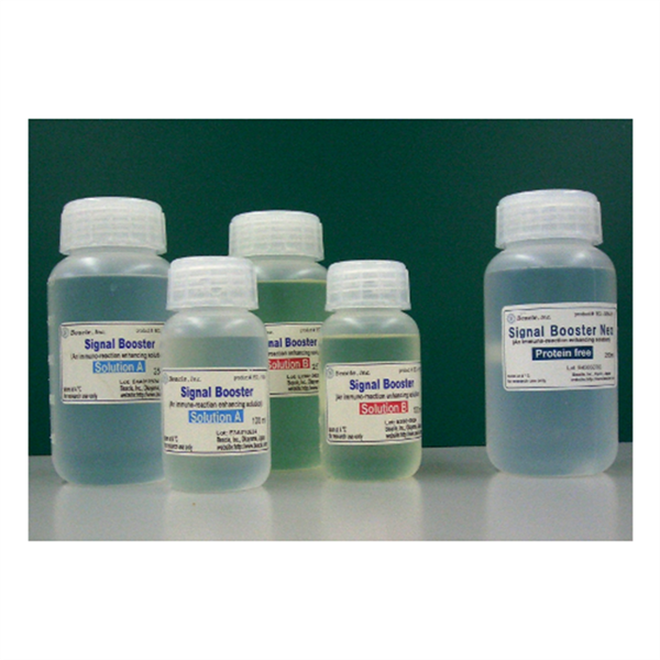
Signal Enhancer
$0.00

Routine PCR
$0.00


Tissue
$0.00
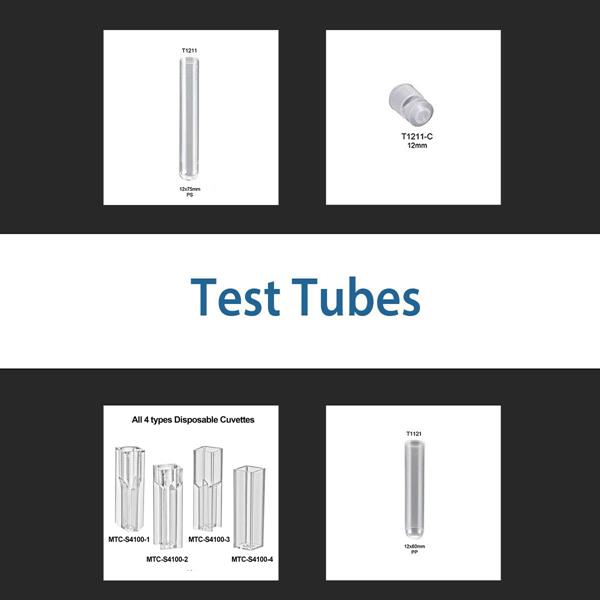
Test Tubes
$0.00
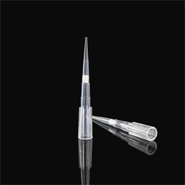
Tips-Non-Sterile/Filter
$0.00
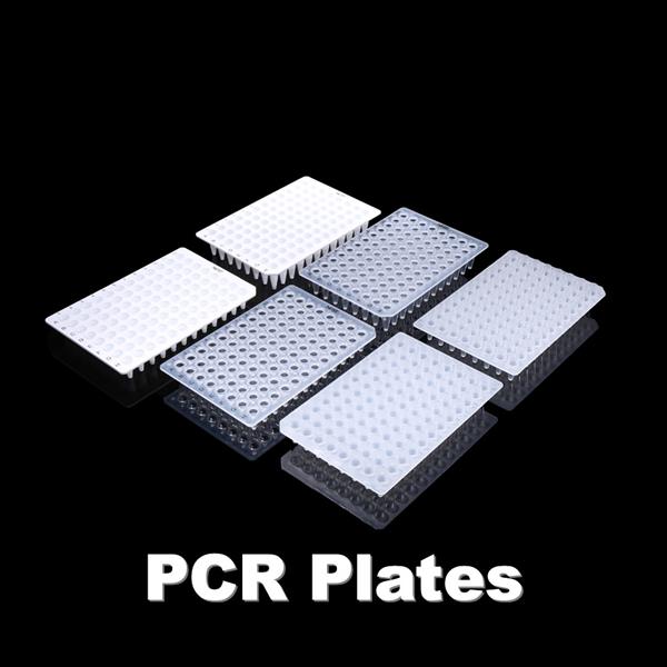
PCR Plates
$0.00

DNA Extract
$0.00

Protein Expression
$0.00

PCR Tubes
$0.00

Fine Scissors
$0.00
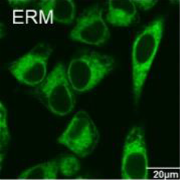
ER-Protein Capture Kit
$717.60
NTP Mix
$635.28

Tube Blocks
$0.00

Dedicated Scissors
$0.00

Dextran
$0.00

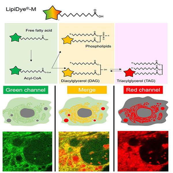
Cellular Imaging
$0.00

Tissue Forceps
$0.00

DNA Tissue
$0.00
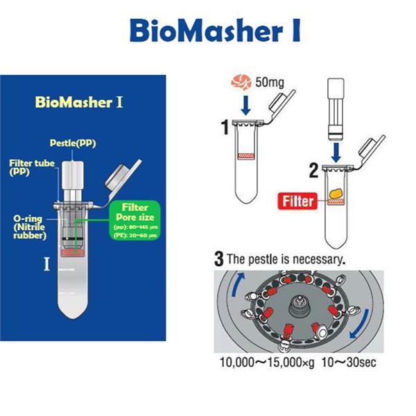
BioMasher I
$0.00
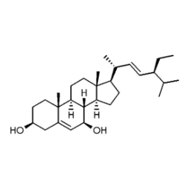
7β-Hydroxystigmasterol
$416.16