

AngioFlamma® TAMRA
Product#: ARR1001
$939.52
Size of product (mg)
-
1 mg

-
5 mg

Availability:
Ships in 1-2 Weeks
AngioFlamma® TAMRA
Cat. No. List below
Description
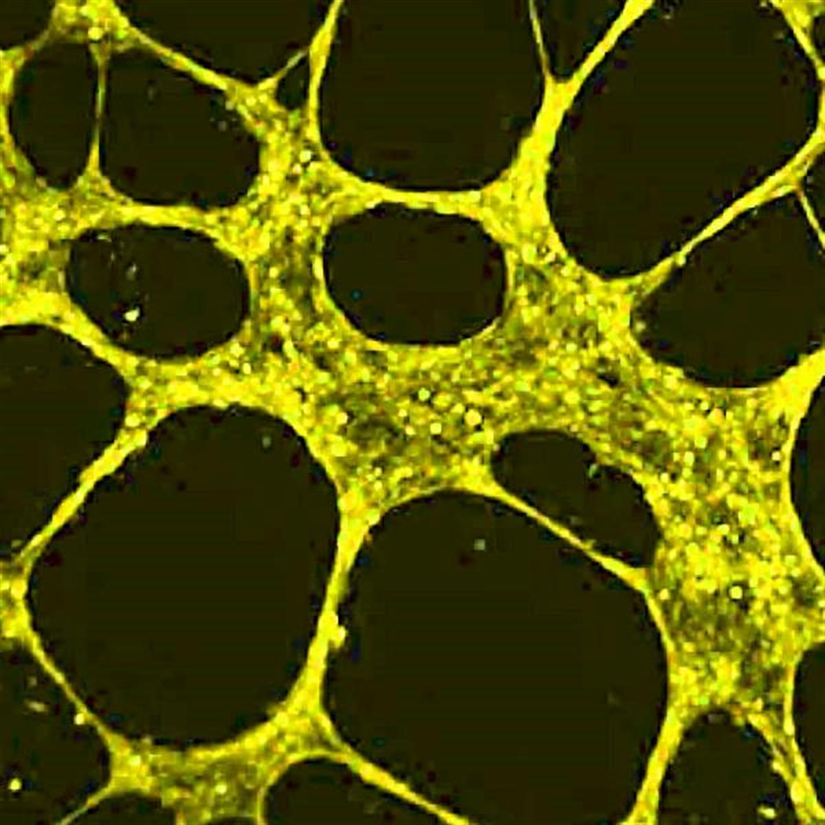
AngioFlamma® TAMRA is an advanced angiogenesis imaging fluorescent probe designed for in vivo visualization of blood vessels, tumors, and angiogenic processes. This innovative probe combines a cyclic RGD (Arg-Gly-Asp) peptide covalently linked to the TAMRA (5(6)-Carboxytetramethylrhodamine) fluorophore, creating a powerful tool for researchers studying vascular biology and tumor progression.
Key Features:
1. Cyclic RGD peptide with high affinity for αvβ3 integrin
2. TAMRA fluorophore with excitation/emission maxima at 543/575 nm
3. Molecular weight of 1032.15 g/mol
4. Red solid appearance
5. Storage at -20°C, protected from light
6. Multi-integrin targeting capability
Applications:
1. In vivo imaging of blood vessels and tumor vasculature
2. Angiogenesis research in cancer biology
3. Evaluation of anti-angiogenic therapies
4. Monitoring integrin expression in various tissues
5. Preclinical drug development and efficacy testing
6. Fluorescence microscopy and flow cytometry studies
Advantages:
1. High specificity: The cyclic RGD peptide binds preferentially to αvβ3 integrin, which is highly expressed in many solid tumors and actively growing blood vessels.
2. Versatile binding: AngioFlamma® TAMRA can interact with multiple integrins (αvβ5, α5β1, α6β4, α4β1, and αvβ6), potentially enhancing tumor uptake and providing a more comprehensive view of the angiogenic process.
3. Sensitive detection: The TAMRA fluorophore offers bright fluorescence in the red spectrum, allowing for sensitive imaging of target structures with minimal background interference.
4. Non-invasive imaging: Enables real-time, non-invasive visualization of angiogenesis and tumor development in living subjects
5. Compatibility: Suitable for use with common fluorescence imaging systems and microscopes equipped with TRITC filter sets.
6. Quantitative analysis: Allows for quantitative assessment of angiogenesis and integrin expression in various tissues and tumors.
7. Stability: TAMRA dye exhibits good photostability, making it suitable for extended imaging sessions and time-lapse studies.
8. Versatility: Can be used for both in vivo imaging and ex vivo analysis of tissue samples.
9. Translational potential: Provides valuable insights into angiogenesis that can be applied to drug development and therapeutic monitoring.
AngioFlamma® TAMRA serves as a valuable tool for researchers and clinicians studying vascular biology, cancer progression, and the efficacy of anti-angiogenic therapies. Its ability to selectively target areas of active angiogenesis, combined with the bright and stable fluorescence of TAMRA, makes it an invaluable asset in the field of molecular imaging and biomedical research. The probe's capacity to bind multiple integrins enhances its utility in capturing a more complete picture of the angiogenic process, potentially leading to new insights in tumor biology and the development of targeted therapies.
Key Features:
1. Cyclic RGD peptide with high affinity for αvβ3 integrin
2. TAMRA fluorophore with excitation/emission maxima at 543/575 nm
3. Molecular weight of 1032.15 g/mol
4. Red solid appearance
5. Storage at -20°C, protected from light
6. Multi-integrin targeting capability
Applications:
1. In vivo imaging of blood vessels and tumor vasculature
2. Angiogenesis research in cancer biology
3. Evaluation of anti-angiogenic therapies
4. Monitoring integrin expression in various tissues
5. Preclinical drug development and efficacy testing
6. Fluorescence microscopy and flow cytometry studies
Advantages:
1. High specificity: The cyclic RGD peptide binds preferentially to αvβ3 integrin, which is highly expressed in many solid tumors and actively growing blood vessels.
2. Versatile binding: AngioFlamma® TAMRA can interact with multiple integrins (αvβ5, α5β1, α6β4, α4β1, and αvβ6), potentially enhancing tumor uptake and providing a more comprehensive view of the angiogenic process.
3. Sensitive detection: The TAMRA fluorophore offers bright fluorescence in the red spectrum, allowing for sensitive imaging of target structures with minimal background interference.
4. Non-invasive imaging: Enables real-time, non-invasive visualization of angiogenesis and tumor development in living subjects
5. Compatibility: Suitable for use with common fluorescence imaging systems and microscopes equipped with TRITC filter sets.
6. Quantitative analysis: Allows for quantitative assessment of angiogenesis and integrin expression in various tissues and tumors.
7. Stability: TAMRA dye exhibits good photostability, making it suitable for extended imaging sessions and time-lapse studies.
8. Versatility: Can be used for both in vivo imaging and ex vivo analysis of tissue samples.
9. Translational potential: Provides valuable insights into angiogenesis that can be applied to drug development and therapeutic monitoring.
AngioFlamma® TAMRA serves as a valuable tool for researchers and clinicians studying vascular biology, cancer progression, and the efficacy of anti-angiogenic therapies. Its ability to selectively target areas of active angiogenesis, combined with the bright and stable fluorescence of TAMRA, makes it an invaluable asset in the field of molecular imaging and biomedical research. The probe's capacity to bind multiple integrins enhances its utility in capturing a more complete picture of the angiogenic process, potentially leading to new insights in tumor biology and the development of targeted therapies.
Specifications
- Fluorophore: TAMRA
- Application: Angiogenesis imaging
- Excitation/Emission Max.(nm): 543/575 nm
- Molecular weight: 1032.15 g/mol
- Appearance: Red Solid
- Storage conditions: -20 ℃, protect from light
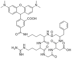
Table 1. List of Annexin V-Flamma® apoptosis detection kits
| Quick link (Cat.#) | Series | λEx (nm) | λEm (nm) | Common filter set | Excitation source | Size |
| ARW1025 | AngioFlamma® FAM | 492 | 519 | FITC | 488 nm | 0.5mg, 1mg, 5mg |
| ARW1011 | AngioFlamma® 552 | 550 | 565 | TRITC | 488, 532 nm | 0.5mg, 1mg, 5mg |
| ARR1001 | AngioFlamma® TAMRA | 543 | 575 | TRITC | 488, 532 nm | 0.5mg, 1mg, 5mg |
| ARW1415 | AngioFlamma® 581 | 581 | 596 | TRITC | 488, 532 nm | 0.5mg, 1mg, 5mg |
| ARW1215 | AngioFlamma® 648 | 648 | 663 | Cy®5 | 594, 633 nm | 0.5mg, 1mg, 5mg |
| ARW1501 | AngioFlamma® 675 | 675 | 691 | Cy®5.5 | 633, 680 nm | 0.5mg, 1mg, 5mg |
| ARW1301 | AngioFlamma® 749 | 749 | 774 | Cy®7 | 680 nm | 0.5mg, 1mg, 5mg |
| ARW1601 | AngioFlamma® 774 | 774 | 806 | Cy®7.5 | 785 nm | 0.5mg, 1mg, 5mg |
| ARO1601 | AngioFlamma® ICG | 785 | 821 | Cy®7.5 | 785 nm | 0.5mg, 1mg, 5mg |
Background
Integrins are heterodimeric transmembrane receptors for cell adhesion to extracellular matrix (ECM) proteins and play important roles in certain cell-cell adhesions. In addition, they make transmembrane connections to the cytoskeleton and activate many intracellular signaling pathways. Because of their role in tumor angiogenesis and progression, integrins have become important diagnostic and therapeutic targets. Integrin αvβ3 is generally expressed in low levels on the epithelial cells and mature endothelial cells, but it is highly expressed in many solid tumors. The αvβ3 levels correlate well with the potential for tumor metastasis and aggressiveness, which make it an important biological target for development of antiangiogenic drugs and molecular imaging probes for early tumor diagnosis. Cyclic tripeptide arginine-glycine-aspartate (RGD) is wellknown to bind preferentially to αvβ3 integrin with high affinity. Cyclic RGD is an effective ligand for tumor targeting since integrin αvβ3 is overexpressed not only on tumoral endothelium but also on various cancer cells. Thus, targeting tumor cells or tumor vasculature by RGD-based strategies is a promising approach for delivering anticancer drugs or diagnostic agents. Cyclic RGD peptides are also able to bind αvβ5, α5β1, α6β4, α4β1, and αvβ6 integrins, which may help enhance their tumor uptake due to the increased receptor population. BioActs developed AngioFlamma® series as effective fluorescent probes for the detection of angiogenesis and tumor cells. The probes are made up of various fluorophores conjugated cyclic RGD. We offer AngioFlamma® series as in vivo fluorescent probes for imaging of blood vessels, tumors and angiogenesis.
Integrins are heterodimeric transmembrane receptors for cell adhesion to extracellular matrix (ECM) proteins and play important roles in certain cell-cell adhesions. In addition, they make transmembrane connections to the cytoskeleton and activate many intracellular signaling pathways. Because of their role in tumor angiogenesis and progression, integrins have become important diagnostic and therapeutic targets. Integrin αvβ3 is generally expressed in low levels on the epithelial cells and mature endothelial cells, but it is highly expressed in many solid tumors. The αvβ3 levels correlate well with the potential for tumor metastasis and aggressiveness, which make it an important biological target for development of antiangiogenic drugs and molecular imaging probes for early tumor diagnosis. Cyclic tripeptide arginine-glycine-aspartate (RGD) is wellknown to bind preferentially to αvβ3 integrin with high affinity. Cyclic RGD is an effective ligand for tumor targeting since integrin αvβ3 is overexpressed not only on tumoral endothelium but also on various cancer cells. Thus, targeting tumor cells or tumor vasculature by RGD-based strategies is a promising approach for delivering anticancer drugs or diagnostic agents. Cyclic RGD peptides are also able to bind αvβ5, α5β1, α6β4, α4β1, and αvβ6 integrins, which may help enhance their tumor uptake due to the increased receptor population. BioActs developed AngioFlamma® series as effective fluorescent probes for the detection of angiogenesis and tumor cells. The probes are made up of various fluorophores conjugated cyclic RGD. We offer AngioFlamma® series as in vivo fluorescent probes for imaging of blood vessels, tumors and angiogenesis.
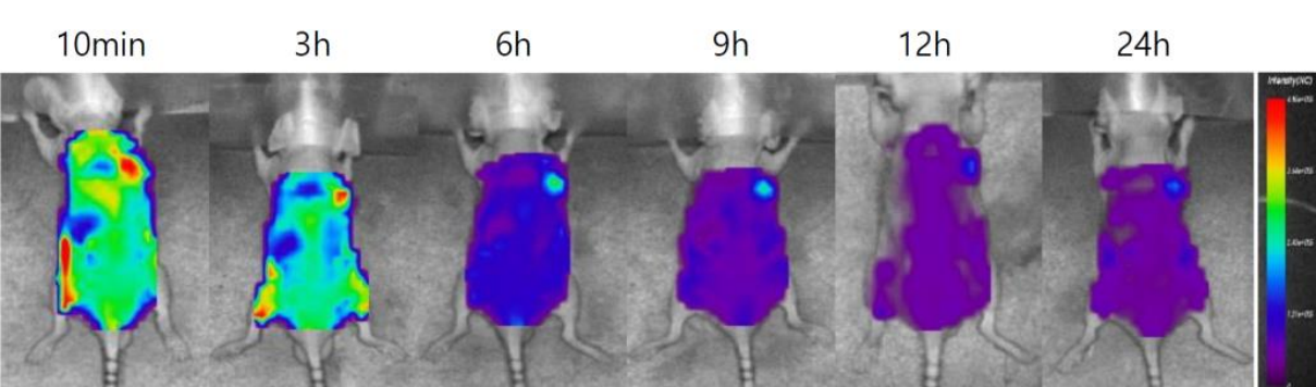
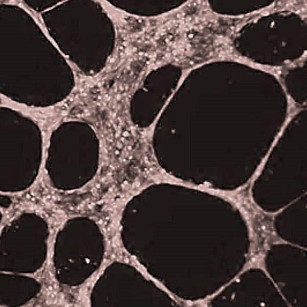
Figure 1. Breast cancer cell imaging using AngioFlamma® 675
In vivo Imaging protocol
1. Preparation of Xenograft tumor model
① MDA-MB-231 human breast cancer cells (1.0 x 10⁷) are injected subcutaneously on the female nude mouse shoulder.
② After the tumor cells have grown to about 50-80 mm3, administer the drug.
2. Injection and Fluorescence imaging
① Fluorescence imaging would be performed up to 24 hours after injection using the optical imaging system.
② Prepared mice is injected intravenously with 100 μg / 200 μL of AngioFlamma® series and images will be taken at appropriate time interval.
① MDA-MB-231 human breast cancer cells (1.0 x 10⁷) are injected subcutaneously on the female nude mouse shoulder.
② After the tumor cells have grown to about 50-80 mm3, administer the drug.
2. Injection and Fluorescence imaging
① Fluorescence imaging would be performed up to 24 hours after injection using the optical imaging system.
② Prepared mice is injected intravenously with 100 μg / 200 μL of AngioFlamma® series and images will be taken at appropriate time interval.
Citation & Reference
- Yumin Zheng. FITC-Conjugated Cyclic RGD Peptides as Fluorescent Probes for Staining Integrin αvβ3/αvβ5 in Tumor Tissues. Bioconjug Chem 25.1 (2014): 1925-41.
- Dongfang Yue. Biomarker-targeted fluorescent probes for breast cancer imaging. Chinese Chemical Letters 29.5 (2018): 648-656.

Recently Viewed
Micro Plate Seal
$0.00

Xyltech H-Fbro
$0.00
Reservoir Plates
$0.00
AngioFlamma® ICG
$939.52
SSPE (20X)
$88.84