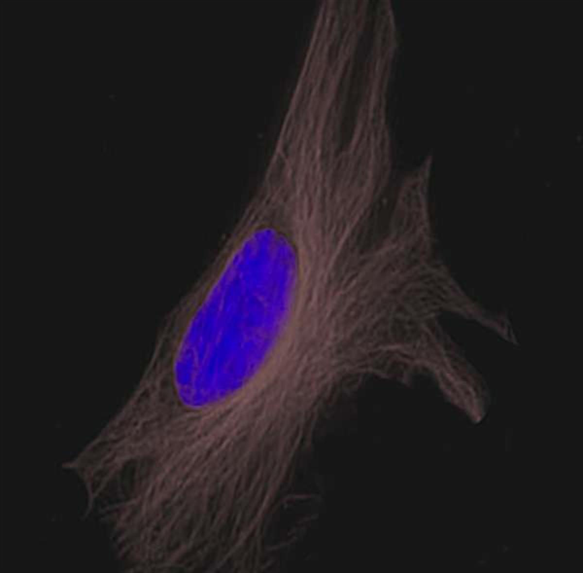
Goat anti-mouse IgG, FSD™ 750
Size of product (mg)
-
0.5 mg

-
1 mg

Goat anti-mouse IgG, FSD™ 750
Cat. No. List below
Description
Key Features:
I. Superior Optical Properties: FSD™ 750, part of the new generation FSD Fluor™ dye series, boasts superior fluorescence intensity and high quantum yield compared to traditional dyes. With excitation and emission maxima at 751 nm and 774 nm respectively, this conjugate is optimized for near-infrared (NIR) imaging.
II. Enhanced Sensitivity: The ability of multiple secondary antibodies to bind to a single primary antibody allows for significant signal amplification. This characteristic enables the visualization of low-abundance targets and potentially reduces experimental time.
III. Versatility: Goat anti-mouse IgG, FSD™ 750 is suitable for a wide range of biological experiments, including:
i. Fluorescence microscopy
ii. Flow cytometry
iii. Microplate assays
iv. Protein and nucleic acid blots
v. In situ hybridization
The Goat anti-mouse IgG, FSD™ 750 is particularly well-suited for applications requiring high sensitivity and low background fluorescence. Its near-infrared spectral properties make it excellent for deep tissue imaging and multiplexing experiments where signal separation is crucial.
What are the advantages?
1. High Specificity: Demonstrates excellent specificity for mouse IgG, ensuring accurate target detection.
2. Signal Amplification: Provides great sensitivity through signal amplification, allowing for detection of even low-abundance targets.
3. Reduced Experimental Time: The high sensitivity may lead to shorter incubation times and overall faster experimental protocols.
4. Low Cross-Reactivity: Minimizes non-specific binding, resulting in cleaner and more reliable results.
5. Compatibility: Optimized for use with 750 nm laser excitation, making it compatible with many advanced imaging systems.
FSD™ 680 is spectrally similar to Alexa Fluor 680 and IRDye 680, but may offer superior performance in terms of brightness and photostability. Its near-infrared emission spectrum makes it an excellent choice for in vivo imaging, deep tissue penetration, and applications where background autofluorescence is a concern.
Goat anti-mouse IgG, FSD™ 750 represents a cutting-edge tool in immunofluorescence detection, offering researchers a powerful combination of sensitivity, specificity, and versatility for their molecular and cellular imaging needs.
Specifications
- Conjugate: FSD™ 750
- Species reactivity: Mouse
- Target: IgG
- Excitation/Emission Max.(nm): 751/774
- Host/Isotype: Goat/IgG
- Type: Secondary antibody
- Antibody form: Whole antibody
- Form: Liquid
- Concentration: 2 mg/mL
- Storage buffer: PBS (pH 7.4)
- Contains: 1.5% BSA, 5 mM sodium azide
- Storage conditions: 4 ℃, protect from light
| Quick link (Cat.#) | Series | Quick link (Cat.#) | Series |
| RSA1145 | Goat anti-mouse IgG, FSD™ 488 | RSA1141 | Goat anti-mouse IgG, Flamma® 488 |
| RSA1155 | Goat anti-mouse IgG, FSD™ 555 | RSA1151 | Goat anti-mouse IgG, Flamma® 552 |
| RSA1195 | Goat anti-mouse IgG, FSD™ 594 | RSA1191 | Goat anti-mouse IgG, Flamma® 594 |
| RSA1165 | Goat anti-mouse IgG, FSD™ 647 | RSA1161 | Goat anti-mouse IgG, Flamma® 648 |
| RSA1175 | Goat anti-mouse IgG, FSD™ 680 | RSA1171 | Goat anti-mouse IgG, Flamma® 675 |
| RSA1105 | Goat anti-mouse IgG, FSD™ 750 | RSA1101 | Goat anti-mouse IgG, Flamma® 749 |
| RSA1185 | Goat anti-mouse IgG, FSD™ 800 | RSA1122 | Goat anti-mouse IgG, HRP |
Background
Fluorescent Protein/Antibody
Fluorescence labeled proteins and antibodies might provide great advantages in the identification of their binding partners or in the observation of biological processes. They allow to visualize cellular environment, to define their function, regulation and interactions. In advanced assays, fluorescent proteins can be utilized in measuring of protein–protein interactions or conformational changes, by measuring intensity changes or FRET energy transfer. An ideal fluorescent label should be small, bright, stable, and without any perturbation to the biological system. Additionally, the fluorescence labeling should be performed at non-active site with the possibility for multiplexing and avoided to the tendency to aggregate.
Organic fluorescent dye-labeled proteins have superior properties over fluorescent protein–conjugated counterparts including, wide spectral range, smaller size, greater photostability, and higher brightness. However, dye–labeled proteins also have some limitations that their sensitivity is low due to lack of the amplification step associated with the secondary antibody, and the protein should be free of contaminants and stabilizing agents. BioActs provides a wide range of fluorophore-conjugated proteins and antibodies for a variety of biological applications.
Fluorescent Dye conjugated Secondary Antibody
Indirect or secondary immunofluorescence method employs specific interactions between primary and secondary antibody. It is more commonly used than the direct immunofluorescence method due to strong signal amplification and cost-effectiveness. Non-labeled primary antibody in indirect immunofluorescence specifically binds to target molecule, and fluorescence conjugated secondary antibody acts as an antigen of the primary antibody. In addition, the polyclonal nature of secondary antibody features multiplex binding of secondary antibodies per a primary antibody resulting in amplified fluorescence signal. However, since indirect immunofluorescence method utilizes complicated antibody-antibody interaction, the experiment process is complicated and the nonspecific fluorescence signals along with cross reaction might occur. BioActs provides a wide spectrum of fluorescent dye conjugated secondary antibodies directed against IgG from mouse, rabbit, rat, and goat.
In spite of competing approaches such as peptide tagging or mass spectrometry, antibody-based detection is the most broadly used application in analysis of specific protein in complex samples. Fluorescent secondary antibodies are raised against IgG heavy and light chains of the target IgG and minimized cross reactivity by affinity purification and by adsorption against the sera of a number of species. For multiple labeling experiments where cross reactivity is the critical issue, we prepare highly cross-adsorbed goat anti–mouse IgG, goat anti–rabbit IgG, and goat anti–rat IgG fluorescent antibodies. Our antibody probes are conjugated with Flamma® Fluor series, which displays strong fluorescence and photostability, along with ICG and horseradish peroxidase (HRP). Due to their outstanding optical and biological properties, Flamma® Fluor conjugates are superior to most conventional fluorescent secondary antibodies and being optimal molecular probes in many applications. BioActs offers series of fluorescent secondary antibodies as suitable molecular probes for many biological experiments such as fluorescence microscopy, flow cytometry, microplate assays, as protein and nucleic acid blots, in situ hybridization, etc.
Citation & Reference
1. Agustina, Lia. Visualization of the physical and functional interaction between hMYH and hRad9 by Dronpa bimolecular fluorescence complementation. BMC molecular biology 15.1 (2014): 1.
2. Lee, Cheol-Jung. Magnolin inhibits cell migration and invasion by targeting the ERKs/RSK2 signaling pathway. BMC cancer 15.1 (2015): 1.
3. Promoter- and RNA polymerase II-dependent hsp-16 gene association with nuclear pores in Caenorhabditis elegans. Rohner S,Kalck V,Wang X,Ikegami K,Lieb JD,Gasser SM,Meister P The Journal of cell biology (200:589)
4. BRAG2/GEP100/IQSec1 interacts with clathrin and regulates α5β1 integrin endocytosis through activation of ADP ribosylation factor 5 (Arf5). Moravec R,Conger KK,D'Souza R,Allison AB,Casanova JE The Journal of biological chemistry (287:31138)