
Flamma® 774 Thiol
Size of product (mg)
-
1 mg

-
5 mg

-
25 mg




Flamma® 774 Thiol
Cat. No. List below
Description
Flamma® Fluors 774 Thiol offers versatile labeling options such as biomolecule labeling (can be attached to biomolecules through disulfide bond formation with thiol groups of cysteine residues), reference standard (useful as a reference standard for dye-conjugates), protein labeling (suitable for protein labeling applications due to its thiol reactivity), in vivo imaging (its NIR properties make it excellent for deep tissue imaging with minimal background interference).
The high extinction coefficient (≥ 182,000 cm?¹M?¹) indicates strong light absorption, contributing to its sensitivity in fluorescence applications.
The low CF280 value (0.1) suggests minimal interference with protein absorbance at 280 nm, making it suitable for protein labeling without significantly affecting concentration measurements.
Its similarity to widely used dyes like Cy7.5 and IRDye800 allows for easy integration into existing imaging protocols and systems.
Flamma® Fluors 774 Thiol represents a powerful tool in the arsenal of fluorescent probes, offering researchers a reliable option for NIR imaging and labeling applications across various fields of life sciences and biomedical research.
What are the advantages of Flamma® Fluors 774 Thiol?
1. Excellent spectral separation with excitation/emission maxima at 774/800 nm, reducing background interference and improving signal-to-noise ratios
2. High extinction coefficient (≥ 182,000 cm?¹M?¹) for strong light absorption and enhanced sensitivity
3. Thiol-reactive nature allows for specific and stable labeling of biomolecules, particularly with cysteine residues
4. Compatible with common excitation sources (750 or 785 nm lasers), enhancing versatility for various imaging setups
5. Spectral similarity to widely used fluorophores (IRDye 800, Cy7.5, CF770) enables easy integration into existing protocols
6. Low CF280 value (0.1) minimizes interference with protein absorbance measurements, ideal for protein labeling
7. High stability and signal-to-noise ratio in the NIR region
8. Particularly suitable for deep tissue imaging and in vivo applications due to minimal autofluorescence and maximum penetration
9. Versatile labeling options, including use as a reference standard for dye-conjugates
- Fluorophore: Flamma® Fluors 774
- Functional group: Thiol
- Excitation/Emission Max.(nm): 774/800
- Spectrally similar dyes: Cy7.5, DyLight800, IRDye800
- Extinction coefficient: ≥ 182,000 cm-1M-1
- CF280: 0.1
- Appearance: Green Solid
- Molecular Weight: 988.20 g/mol
- Solubility: DMF, DMSO
- Storage conditions: -20 ℃, protect from light
- Excitation Sources: Can be excited using 750 or 785 nm laser lines or dye-pumped laser excitation
- Emission: Occurs in the NIR region
| Quick link (Cat.#) | FSD series | EXmax (nm) | EMmax (nm) | Spectrally similar dyes |
| CWT1001 | Flamma® 496 Thiol | 494 | 520 | Alexa488, FITC, Cy2 |
| CWT1058 | Flamma® 552 Thiol | 550 | 564 | Alexa555, DyLight549, Cy3, ATTO550 |
| KWT1415 | Flamma® 581 Thiol | 578 | 593 | Alexa594, DyLight594 |
| KWT1042 | Flamma® 648 Thiol | 648 | 663 | Alexa647, DyLight650, Cy5 |
| PWT1415 | Flamma® 675 Thiol | 674 | 691 | Alexa680, DyLight680, Cy5.5, IRDye680LT, CF680 |
| PWT1215 | Flamma® 749 Thiol | 749 | 774 | Alexa750, DyLight755, Cy7.5, IRDye750 |
| PWT1515 | Flamma® 774 Thiol | 774 | 800 | Cy7.5, DyLight800, IRDye800 |
Background
Flamma® Fluors
BioActs offers a broad range of Flamma® Fluors dyes equipped with variety of reactive and functional groups, which can cover the full spectral range from UV to NIR with their excellent fluorescence performance. Characteristic features of these superior dyes are strong absorption, high fluorescence quantum yield and high photostability. Flamma® dyes maintain good fluorescence activity and stability after conjugation to biomolecules and allow the detection of low-abundance biological structures with great sensitivity. The dyes are compatible with optical conditions of most of fluorescent equipment and are ideal for any applications in biological studies.
- Covering the full spectral range from UV to NIR
- Equipped with a variety of reactive groups: NHS and Sulfo-NHS ester, Vinylsulfone, Maleimide, Click chemistry, isothiocyanate, hydrazide and hydrophobic substances.
- High quantum yields and photostability
- High purity and compatible with most of biomolecules
Flamma® Fluors Thiol
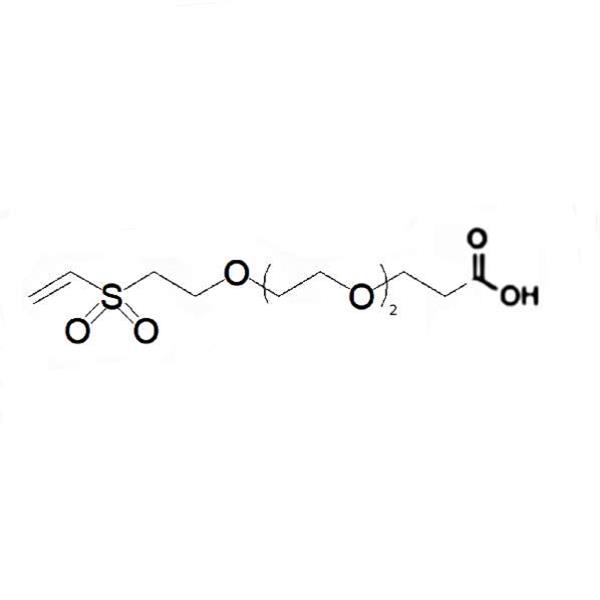
Flamma® Fluors thiol dyes have an attached primary thiol, which connected through a spacer. These thiol dyes can be labeled to biomolecules through disulfide bond formation with thiol of cysteine residue.

Figure 1. Absorption (upper) and emission (bottom) spectra overlap of Flamma® Fluors

Figure 2. Immunofluorescence imaging and in situ hybridization imaging

Figure 3. Fluorescence images of Flamma® 749 (upper) and Flamma® 774 (bottom) carboxylic acid injected mouse model
Citation & Reference
1. Xu, Peisheng. Zwitterionic chitosan derivatives for pH-sensitive stealth coating. Biomacromolecules 11.9 (2010): 2352-2358.
2. Ibrahim, Basma M. A strategy to deliver genes to cystic fibrosis lungs: a battle with environment. Journal of controlled release 155.2 (2011): 289-295.
3. Oh, Keun Sang. Accurate sequential detection of primary tumor and metastatic lymphatics using a temperature-induced phase transition nanoparticulate system. International journal of nanomedicine 9 (2014): 2955.
4. Yhee, Ji Young. Tumor-targeting transferrin nanoparticles for systemic polymerized siRNA delivery in tumor-bearing mice. Bioconjugate chemistry 24.11 (2013): 1850-1860.
5. Yoon, Hong Yeol. Glycol chitosan nanoparticles as specialized cancer therapeutic vehicles: Sequential delivery of doxorubicin and Bcl-2 siRNA. Scientific reports 4 (2014).
6. Ryu, Ju Hee. Early diagnosis of arthritis in mice with collagen?induced arthritis, using a fluorogenic matrix metalloproteinase 3–specific polymeric probe. Arthritis & Rheumatism 63.12 (2011): 3824-3832.
7. Hollis, Christin P. In vivo investigation of hybrid paclitaxel nanocrystals with dual fluorescent probes for cancer theranostics. Pharmaceutical research 31.6 (2014): 1450-1459.
8. Koo, Heebeom. The movement of self-assembled amphiphilic polymeric nanoparticles in the vitreous and retina after intravitreal injection. Biomaterials 33.12 (2012): 3485-3493.
9. Zhu, Lei. Real-time monitoring of caspase cascade activation in living cells. Journal of controlled release 163.1 (2012): 55-62.
10. Yoon, Hong Yeol. Bioreducible hyaluronic acid conjugates as siRNA carrier for tumor targeting. Journal of Controlled Release 172.3 (2013): 653-661.
11. Yhee, Ji Young. Cancer-targeted MDR-1 siRNA delivery using self-cross-linked glycol chitosan nanoparticles to overcome drug resistance. Journal of Controlled Release 198 (2015): 1-9.
12. Park, Jin Woo. Wide-Ranged Fluorescent Molecular Weight Size Markers for Electrophoresis. Bulletin of the Korean Chemical Society 34.1 (2013): 29-30.
13. Huang, Xinglu. Multiplex Imaging of an Intracellular Proteolytic Cascade by using a Broad?Spectrum Nanoquencher. Angewandte Chemie International Edition 51.7 (2012): 1625-1630.