Dextran (10K), Flamma® 774
Dextran (10K), Flamma® 774
Cat. No. RSC0314
Description
Dextran (10K) Flamma® 774 is employed in a broad range of biological investigations, including neural tracing (anterograde and retrograde), cell lineage tracing in live cells, tracking endocytosis. examining intercellular communication
Studying vascular permeability
Investigating blood-brain barrier integrity
Analyzing fluid-phase endocytosis
What are the advantages?
1. Long-distance transport: Can be transported over extended distances within biological systems
2. Fixation compatibility: Suitable for use in cut and fixed nerve and brain tissues
3. Versatility: Applicable to a wide range of biochemical and biomedical studies
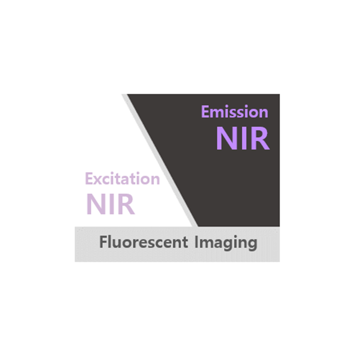
4. Near-infrared spectrum: The Flamma® 774 fluorophore's excitation and emission wavelengths fall within the near-infrared range, allowing for deeper tissue penetration and reduced autofluorescence
5. Low toxicity: Dextrans are known for their low toxicity and good biocompatibility
Dextran (10K) Flamma® 774 is typically introduced into cells via microinjection. Its relatively large size (10K) allows for longer retention within cells compared to smaller dextrans, making it ideal for extended studies. The fluorescent label enables researchers to track its movement and distribution within biological systems, providing valuable insights into cellular processes and tissue dynamics.
Dextrans are hydrophilic polysaccharides with good water solubility. They are biologically inert due to their uncommon poly-(α-D-1,6-glucose) linkages, which render them resistant to cleavage by most endogenous cellular glycosidases. Flamma® 774 is comparable to other near-infrared dyes such as Cy7.5, IRDye 800, and CF770. The dye can be excited using a 633 nm laser line or dye-pumped laser excitation.
Dextran (10K) Flamma® 774 is a powerful and versatile tool for researchers in cell biology, neuroscience, and related fields, offering high-quality fluorescent labeling and compatibility with a wide range of experimental protocols and imaging techniques.
Specifications
- Fluorophore: Flamma® 774
- Molecular Weight: 10,000 daltons
- Application: Neural tracing, cell lineage tracing, tracking endocytosis
- Excitation/Emission Max.(nm): 774/806 nm
- Absorption λ max: 774 nm
- Emission λ max: 806 nm
- Storage conditions: 4℃, protect from light
- Molar Extinction Coefficient (ε max): 182,000 M?¹ cm?¹
Dextran
| Quick link (Cat.#) | Series | Excitation | Emission |
| RSC0302 | Dextran (10K), Flamma® 488 | 495 | 519 |
| RSC0305 | Dextran (10K), Flamma® 552 | 550 | 565 |
| RSC0306 | Dextran (10K), Flamma® 560 | 560 | 589 |
| RSC0310 | Dextran (10K), Flamma® 581 | 581 | 596 |
| RSC0311 | Dextran (10K), Flamma® 648 | 650 | 665 |
| RSC0312 | Dextran (10K), Flamma® 675 | 675 | 691 |
| RSC0313 | Dextran (10K), Flamma® 749 | 749 | 774 |
| RSC0314 | Dextran (10K), Flamma® 774 | 774 | 806 |
Background
Biochemical Polymers & Labeling
The optimal labeling condition for biomolecules is to achieve an appropriate degree of conjugation ratio yet to retain the important functionality of the original biomolecules such as binding affinity, activatory or inhibitory activity, solubility and biological membrane permeability. The high-number of labeling often causes conjugated biomolecule to precipitate out of solution or to lose its functional properties, thus the degree of labeling should be determined from experimental optimization process. There are two major types of reactive dyes: amine-reactive dye and thiol-reactive dye. The primary target of amine-reactive probes at protein is lysine residue, and thiol residue is the main target for thiol-reactive probes. In mammalian proteins, the occurrence frequency of lysine residue is 7.2% and that of thiol is 3.3%.
Amino-labeling is the widely utilized method to conjugate proteins, peptides, oligonucleotides and other biomolecules with dyes. Amine-reactive dyes might be used to prepare bioconjugates for fluorescent analog cytochemistry, immunochemistry, cell tracing, receptor labeling, FITC, etc. The primary target for amine-reactive probe is lysine residue, which has the fifth highest occurrence frequency of the 20 natural amino acids in mammalian proteins. A typical IgG antibody has about 90 lysine residues, and the maximum number of labeling will be around 30 residues with excess amount of reagent and prolonged incubation. However, maintaining functional properties requires the degree of labeling less than 10 dyes per antibody. BioActs provides three major classes (NHS, Sulfo-MHS and Vinylsulfone) of amine-reactive Flamma® Fluors dyes, and they can cover the entire spectral range from visible to NIR region.
Thiol-reactive dyes are mainly used for labeling proteins for the observation of conformational change, multi-subunit complexes assembly and ligand-binding processes. In proteins and peptides, the primary targets of thiol-reactive probes are cysteine residues. Unlike amine-labeling, the low abundance of cysteine residues enable to achieve saturated labeling without risk of conjugated protein precipitation and fluorescence self-quenching interactions. Thiols play a principal role in maintaining the appropriate oxidation–reduction state of proteins, cells and organisms, and they are easily oxidized to form disulfides. Thiols can also be generated by the reduction of cysteine disulfides with reducing agents such as dithiothreitol (DTT), 2-mercaptoethanol or tris-(2-carboxyethyl)phosphine (TCEP). However, the reducing process may cause to disrupt the tertiary structure of protein. Maleimide is a well-known reactive group that can specially label thiol of cysteine residue without interacting with amino functionality. In labeling process, thiol is added to the double bond of maleimide via 1,4-addition pathway to form thioether linkage. Maleimides apparently do not react with methionine, histidine or tyrosine, but they also react with amines in the strong basic environment. BioActs offers Flamma® Fluors maleimide series as thiol-reactive fluorescence dyes.
Click chemistry is a typical type of bioorthogonal reactions, which the reaction occurs inside of living systems yet without interfering with native biochemical processes. The most widely utilized click chemistry is 1,3-dipolar cycloaddition between an azide and an alkyne to produce 1,4-disubstituted 1,2,3-triazole. The triazole ring is stable under hydrolysis, oxidation or reduction, and it survives ionization process in mass spectrometry (MS) analysis. There are two types of 1,3-dipolar cycloaddition methods: copper(I)-catalyzed azide-alkyne cycloaddition (CuAAC) and strain-promoted azide-alkyne cycloaddition (SPAAC). BioActs offers Flamma® Fluors Alkyne dyes for CuAAC, Flamma® Fluors ADIBO products for SPAAC and Flamma® Fluors Azide dyes for both CuAAC and SPAAC.
Fluorescent Dextran
Dextrans are hydrophilic water soluble polysaccharides that have been used in a wide variety of biomedical applications. Characteristics of dextran are moderate to high molecular weight, good water solubility and low toxicity. Dextrans are biologically inert due to their uncommon poly-(α-D-1,6-glucose) linkages, which render them resistant to cleavage by most endogenous cellular glycosidases. Dextrans might be used as either anterior or retro tracers depending on the experiment protocol and tissue type used. Fluorescence labeled dextrans are routinely employed in microscopic studies to monitor cell division, to trace neuronal projections and track the movement of live cells, and to report the hydrodynamic properties of the cytoplasmic matrix.
The labeled dextran is commonly introduced into the cells via microinjection. Due to widely variable size and low toxicity, fluorescent dextran conjugates are utilized in a variety of biological studies such as cell lineage tracing in live cells, intercellular communication examination, vascular permeability, tracking endocytosis, and blood–brain barrier integrity. BioActs developed two types of fluorescence conjugated dextrans: Dextran (3K) and (10K) series. Dextran (3K) series is a wide range of fluorescent dyes conjugated 3000 dalton dextran, and in dextran (10K), the size of dextran is 10000 dalton. The 3K dextrans display several advantages over higher size dextrans, including faster axonal diffusion and greater access to peripheral cell processes. However, dextran (10K) can be transported over longer distance and can be applied to cut and fix nerves and brain tissues. Dextran 3K and 10K might be utilized in a range of biochemical and biomedical applications, including for analyzing fluid-phase endocytosis, for tracing cell lineage, for examining intercellular communications, and for investigating vascular permeability and blood–brain barrier integrity.
Citation & Reference
1. DWIGHT M. NANCE. Fluorescent Dextrans as Sensitive Anterograde Neuroanatomical Tracers: Applications and Pitfalls. Brain Res Bull 25.1 (1990): 139-45.
2. Anne-Marie Ellegaard. Visualizing Lysosomal Membrane Permeabilization by Fluorescent Dextran Release. Cold Spring Harb Protoc 2015.10 (2015): 900-3.
3. M.J. Dolleman-Van der Weel. Multiple anterograde tracing, combining Phaseolus vulgaris leucoagglutinin with rhodamine- and biotin-conjugated dextran amine. J Neurosci Methods 51.1 (1994): 9-21.