UltraRIPA kit for Lipid Raft (1 kit)
UltraRIPA kit for Lipid Raft
Cat. No. FNK-F015
Size 1 kit
NIH NSN# 664000N172813
Kit components
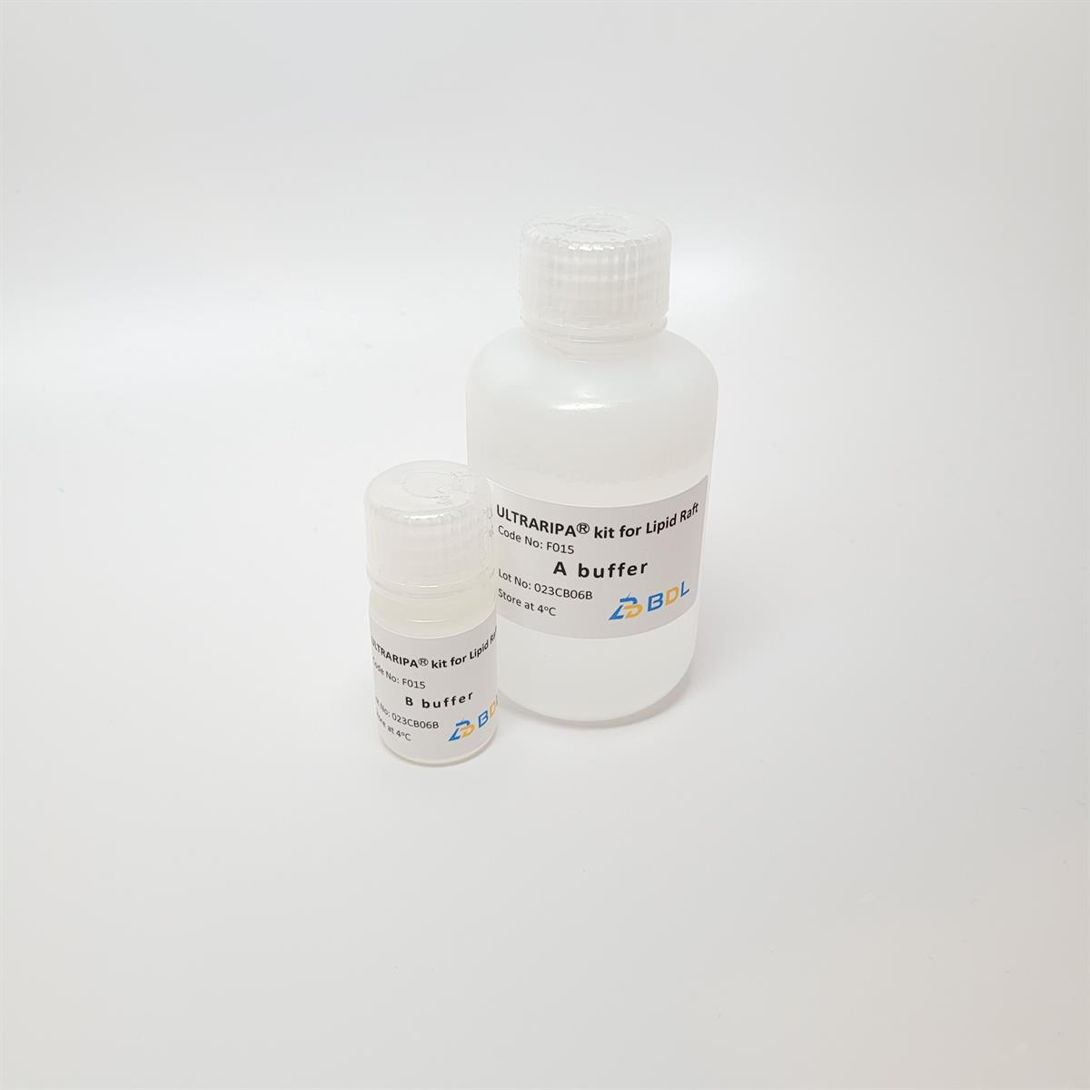
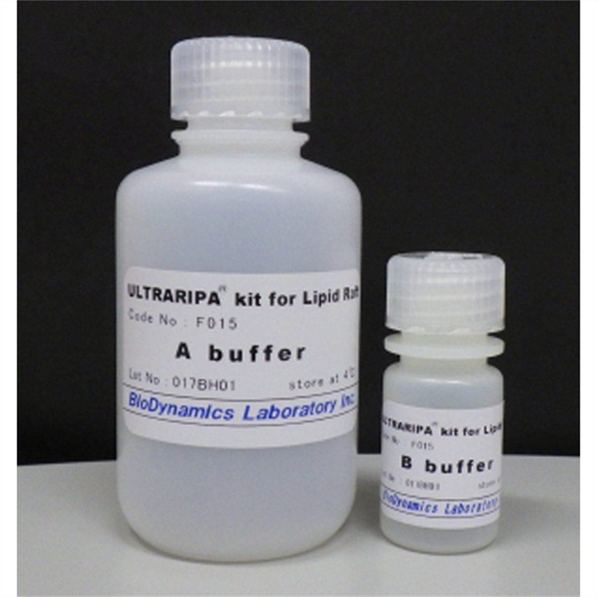
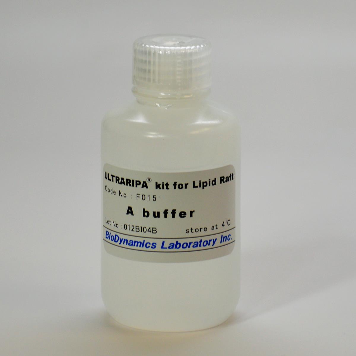
A buffer (RIPA buffer) 100 mL
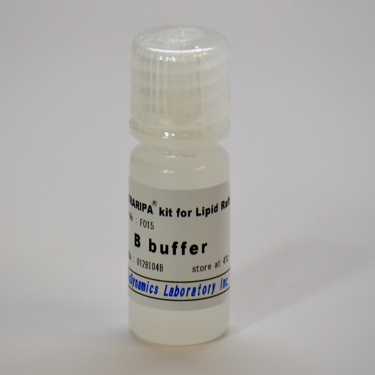
B buffer 10 mL
Store at 4oC, Product shelf life 1 year
Product ID and Size
| Cat No. | Size | Link |
| FNK-F015 | 1 Kit |  |
| FNK-F015-Trial | 1 Kit | |
Description
Next-Generation RIPA Buffer for High Efficient Membrane Protein Extraction
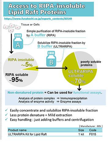
RIPA buffer is one of the most useful buffers for protein extraction. RIPA buffer maintains most native structures of proteins and the extracted proteins can be applied to various applications. However, RIPA buffer is not sufficient to extract membrane proteins and membrane-associated proteins concentrated in lipid raft.
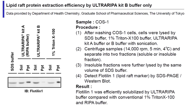
Lipid raft is a highly specialized microdomain on the lipid bilayer which contains specialized lipids, cholesterol and functional proteins. These lipid rafts are also called “Detergent Resistant Membrane (DRM)”, as lipid raft-enriched proteins are usually insoluble by mild detergent buffers such as 1% Triton X-100 and RIPA buffer. Consequently, it was difficult to analyze functions of lipid raft-enriched proteins extracted with RIPA buffer.
BioDynamics Laboratory's newly developed product : the UltraRIPA kit, can efficiently and rapidly extract membrane proteins or membrane-associated proteins enriched in lipid rafts with native structure and function.
UltraRIPA kit includes a totally new buffer not containing protein denaturing detergents, but can extract the DRM which was difficult to extract by conventional RIPA buffer. UltraRIPA kit helps to analyze various biological assays of the proteins in lipid raft.
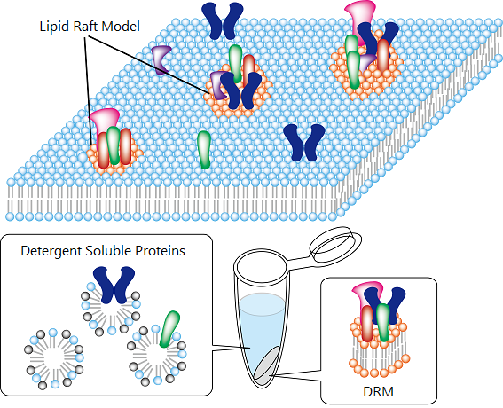
Features of UltraRIPA kit
- Extract membrane / membrane associated proteins enriched in lipid rafts, with native structure and fully retained function.
- Easy and Simple Procedure : Only a centrifuge is required
- Only two components: A buffer and B buffer
Overview of Procedure
Kit components
A buffer (RIPA Buffer) : 100 mL
B buffer : 10 mL
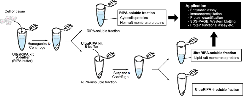
Overview of advantages of ULTRARIPA kit
|
Protein Extraction Buffer |
Protein Extraction |
Protein Structure |
Protein Function |
Application |
||
|
Cytosolic |
Membrane |
|||||
|
Non-lipid raft |
Lipid raft |
|||||
|
> 1% SDS buffer |
|
|
|
|
|
SDS-PAGE |
|
RIPA buffer |
|
|
|
|
|
Enzymatic assay, |
|
ULTRARIPA kit |
|
|
|
|
|
|


Protein extraction efficiency of ULTRARIPA kit
 |
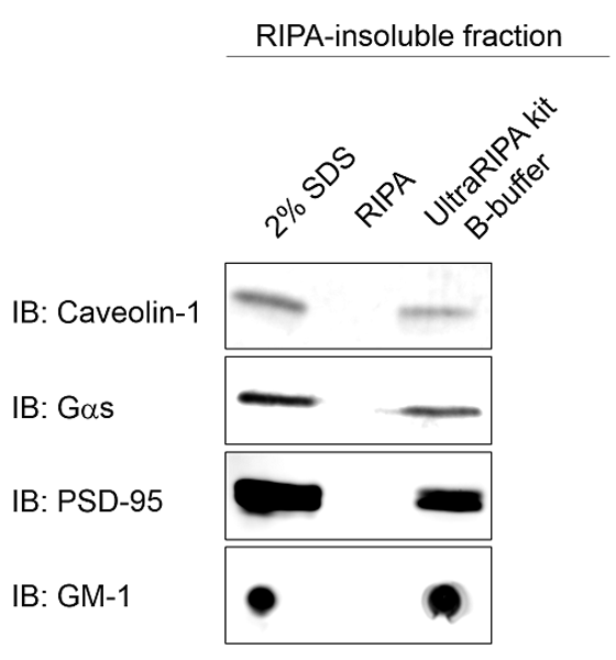 |
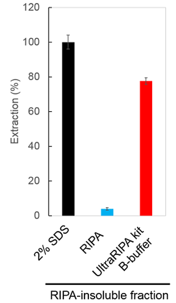
Extraction of RIPA-insoluble proteins by ULTRARIPA kit
Left : Quantification of extracted total proteins by BCA protein assay. ULTRARIPA kit could constantly extract over 70% of RIPA-insoluble proteins from the mouse brain tissue.
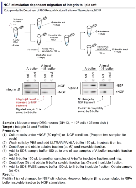
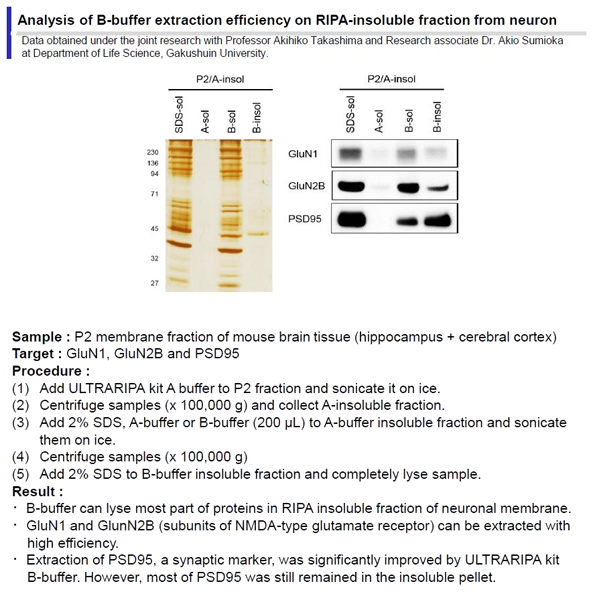
Right : Western blotting of lipid raft markers. Some lipid raft markers among proteins or a ganglioside extracted were dramatically increased in RIPA-insoluble fraction by ULTRARIPA kit.
Enzyme activity assay of proteins extracted by ULTRARIPA kit
Left : Lactate Dehydrogenase activity of proteins extracted by using 1% Triton X-100, A and B buffer. Equivalent enzyme activity are obtained when using A and B buffer.
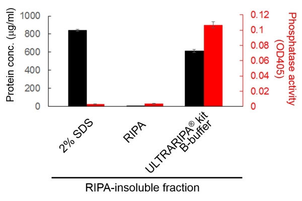
Right: Total protein phosphatase activity. Protein extracts from RIPA-insoluble fraction of the mouse whole brain by ULTRARIPA® kit B-buffer, RIPA, and 2% SDS buffer were applied to total protein phosphatase assay. Although 2% SDS buffer completely extracted proteins, but it disrupted phosphatases. In contrast, ULTRARIPA® kit showed greatly higher activity of protein phosphatases than 2% SDS and RIPA buffer
Note
- Both A and B-buffer could not be applied to Bradford protein assay.
- Please use BCA protein assay if you would like to quantitate protein concentration.
|
Problem |
Possible Cause |
Solution |
|
Low RIPA-insoluble fraction yield |
Less of total protein Use more |
starting cells or tissues |
|
Low concentration of |
Excess buffer used |
proteins Use less buffer |
|
Degradation of proteins |
No protease inhibitors added |
Add any protease inhibitors to the both buffers before use |
Application
Reference
- Taruno A. et al., Post-translational palmitoylation controls the voltage gating and lipid raft association of CALHM1 channel. J Physiol. (2017) 595(18):6121-6145.
- Tan L. et al., Enriched expression of neutral sphingomyelinase 2 in the striatum is essential for regulation of lipid raft content and motor coordination. Mol Neurobiol. (2018) 55(7):5741-5756.
- Toyoda Y. et al., Extracellular glucose level regulates dependence on GRP 78 for cell surface localization of multipass transmembrane proteins in HeLa cells.FEBS Lett. (2018) 592(19):3295-3304.
- Araki K. et al., Mitochondrial protein E2F3d, a distinctive E2F3 product, mediates hypoxia-induced mitophagy in cancer cells. Commun Biol. (2019) 2: 3.
- Hayakawa K. et al., MicroRNA-766-3p Contributes to Anti-Inflammatory Responses through the Indirect Inhibition of NF-κB Signaling. Int J Mol Sci. (2019) 20(4): 809.
- Ding X. et al., Docosahexaenoic Acid Serving As Sensitizing Agents And Gefitinib Resistance Revertants In EGFR Targeting Treatment. Onco Targets Ther. (2019) 12: 10547–10558.
- Linden JR. et al., Clostridium perfringens epsilon toxin induces blood brain barrier permeability via caveolae-dependent transcytosis and requires expression of MAL. PLoS Pathog (2019)15(11):e1008014.
- Tsuruoka K. et al., Skin characteristics associated with foot callus in people with diabetes: A cross-sectional study focused on Desmocollin1 in corneocytes. J Tissue Viability (2020) S0965-206X(20)30077-2.
- Dewa KI et al., Neuronal DSCAM regulates the peri-synaptic localization of GLAST in Bergmann glia for functional synapse formation. Nat Commun. 2024 15(1):458.
- Tumpara S. et al., Polymerization of misfolded Z alpha-1 antitrypsin protein lowers CX3CR1 expression in human PBMCs. 2021. eLife. 2021; 10: e64881.
- Stern AM. et al., Abundant Aβ fibrils in ultracentrifugal supernatants of aqueous extracts from Alzheimer's disease brains. Neuron 2023 111(13):2012-2020.
- Qiu Y. et al., ACSL4-Mediated Membrane Phospholipid Remodeling Induces Integrin β1 Activation to Facilitate Triple-Negative Breast Cancer Metastasis. Cancer Res. 2024 84(11):1856-1871.
- Liu D., Targeted disruption of mitochondria potently reverses multidrug resistance in cancer therapy. Br J Pharmacol. 2022 179(13):3346-3362.
- Tumpara S., Polymerization of misfolded Z alpha-1 antitrypsin protein lowers CX3CR1 expression in human PBMCs.Elife. 2021 18:10:e64881.
- Wang, R., Hao, M., Kou, X., Sui, B., Sanmillan, M. L., Zhang, X., ... & Shi, S. (2022). Apoptotic vesicles ameliorate lupus and arthritis via phosphatidylserine-mediated modulation of T cell receptor signaling. Bioactive Materials, 25, 472–484.
- Chean, J., Chen, C. J., Gugiu, G., Wong, P., Cha, S., Li, H., ... & Shively, J. E. (2021). Human CEACAM1-LF regulates lipid storage in HepG2 cells via fatty acid transporter CD36. Journal of Biological Chemistry, 297(5), 101311.
- Yang, Q., Pei, R., Wang, Y., Zhou, Y., Yang, M., Chen, X., & Chen, J. (2021). ADAM15 Participates in Tick-Borne Encephalitis Virus Replication. Journal of Virology, 95(4), e01926-20.
- Dewa K, Arimura N, Kakegawa W, et al. Neuronal DSCAM regulates the peri-synaptic localization of GLAST in Bergmann glia for functional synapse formation. Nature Communications. 2024;15(1):458. doi:10.1038/s41467-023-44579-z
- Araki K, Kawauchi K, Sugimoto W, et al. Mitochondrial protein E2F3d, a distinctive E2F3 product, mediates hypoxia-induced mitophagy in cancer cells. Communications Biology. 2019;2:3. doi:10.1038/s42003-018-0246-9
- Itakura M, Yamaguchi K, Kitazawa R, et al. Histone functions as a cell-surface receptor for AGEs. Nature Communications. 2022;13:2974. doi:10.1038/s41467-022-30626-8
- Otsuka R, Kajikuri J, Matsui M, et al. LRRC8A Inhibition Overcomes Chemoresistance by Downregulating MRP3 and CYP3A4 in the 3D Spheroid Model of Human Breast Cancer Cells. International Journal of Molecular Sciences. 2026;27(6):2646. doi:10.3390/ijms27062646
- Tsurudome N, Minami Y, Kajiya K. Sphingosylphosphorylcholine (SPC), a Causative Factor of SPC-Induced Vascular Smooth Muscle Cells Contraction, Is Taken Up via Endocytosis. Cells. 2023;12(2):265. doi:10.3390/cells12020265
- Liu S, Tao J, Duan F, Li H, Tan H. HHcy Induces Pyroptosis and Atherosclerosis via the Lipid Raft-Mediated NOX-ROS-NLRP3 Inflammasome Pathway in apoE−/− Mice. Cells. 2022;11(15):2438. doi:10.3390/cells11152438
- Murae M, Shimizu Y, Yamamoto Y, et al. The function of SARS-CoV-2 spike protein is impaired by disulfide-bond disruption with mutation at cysteine-488 and by thiol-reactive N-acetyl-cysteine and glutathione. Biochemical and Biophysical Research Communications. 2022;597:30–36. doi:10.1016/j.bbrc.2022.01.106
- Yamamoto Y, Nakano Y, Murae M, et al. Direct Inhibition of SARS-CoV-2 Spike Protein by Peracetic Acid. International Journal of Molecular Sciences. 2022;24(1):20. doi:10.3390/ijms24010020
- Hayashi T, Yamamoto N, Kurosawa G, et al. A Novel High-Throughput Screening Method for a Human Multicentric Osteosarcoma-Specific Antibody and Biomarker Using a Phage Display-Derived Monoclonal Antibody. Cancers (Basel). 2022;14(23):5829. doi:10.3390/cancers14235829
- Mai S, Izumi K, Mai Y, et al. Native Autoantigen Complex Detects Pemphigoid Autoantibodies. JID Innovations. 2023;3(3):100193. doi:10.1016/j.xjidi.2023.100193
- Yokota K, Yamada H, Mori H, et al. Platelet-Derived Growth Factor Subunit A Strengthens the Neurovascular Unit and Inhibits Retinal Vascular Regression Under Hyperoxic Conditions. International Journal of Molecular Sciences. 2024;25(23):12945. doi:10.3390/ijms252312945
- Chan NN, Yamazaki M, Maruyama S, et al. Cholesterol Is a Regulator of CAV1 Localization and Cell Migration in Oral Squamous Cell Carcinoma. International Journal of Molecular Sciences. 2023;24(7):6035. doi:10.3390/ijms24076035
- Ohya S, Kajikuri J, Endo K, Kito H, Matsui M. KCa1.1 K+ Channel Inhibition Overcomes Resistance to Antiandrogens and Doxorubicin in a Human Prostate Cancer LNCaP Spheroid Model. International Journal of Molecular Sciences. 2021;22(24):13553. doi:10.3390/ijms222413553
- Umemoto K, Nakamura T, Sasaki K, et al. Platelets and MMP-9 contribute to esophageal cancer invasion via CD40-CD154 interactions. Oncology Reports. 2025;54(1):79. doi:10.3892/or.2025.8912
- Takai Y, Naito S, Ito H, et al. Ankrd1 Promotes Lamellipodia Formation and Cell Motility via Interaction with Talin-1 in Clear Cell Renal Cell Carcinoma. International Journal of Molecular Sciences. 2025;26(9):4232. doi:10.3390/ijms26094232
- Tahara M, Higurashi N, Hata J, et al. Developmental changes in brain activity of heterozygous Scn1a knockout rats. Frontiers in Neurology. 2023;14:1125089. doi:10.3389/fneur.2023.1125089
- Ito S, Hashimoto H, Yamakawa H, et al. The complement C3-complement factor D-C3a receptor signalling axis regulates cardiac remodelling in right ventricular failure. Nature Communications. 2022;13(1):5409. doi:10.1038/s41467-022-33152-9
- Frankish J, Mukherjee D, Romano E, et al. The CD40 agonist HERA-CD40L results in enhanced activation of antigen presenting cells, promoting an anti-tumor effect alone and in combination with radiotherapy. Frontiers in Immunology. 2023;14:1160116. doi:10.3389/fimmu.2023.1160116
- Ohya S, Kajikuri J, Endo K, et al. Ca2+?activated K+ channel KCa1.1 as a therapeutic target to overcome chemoresistance in three?dimensional sarcoma spheroid models. Cancer Science. 2021;112(9):3769–3783. doi:10.1111/cas.15046
- Zhao X, Zhao Z, Li B, et al. ACSL4-mediated lipid rafts prevent membrane rupture and inhibit immunogenic cell death in melanoma. Cell Death & Disease. 2024;15(9):695. doi:10.1038/s41419-024-07098-3
- Chen LJ, Zhang NN, Zhou CX, et al. Gm364 coordinates MIB2/DLL3/Notch2 to regulate female fertility through AKT activation. Cell Death & Differentiation. 2022;29(2):366–380. doi:10.1038/s41418-021-00861-5
- Kabwe JC, Sawada H, Mitani Y, et al. CRISPR-mediated Bmpr2 point mutation exacerbates late pulmonary vasculopathy and reduces survival in rats with experimental pulmonary hypertension. Respiratory Research. 2022;23(1):87. doi:10.1186/s12931-022-02005-w
- Taruno A, Sun H, Nakajo K, et al. Post-translational palmitoylation controls the voltage gating and lipid raft association of the CALHM1 channel. The Journal of Physiology. 2017;595(18):6121–6145. doi:10.1113/JP274164
- Im E, Jiang Y, Stavrides PH, et al. Lysosomal dysfunction in Down syndrome and Alzheimer mouse models is caused by v-ATPase inhibition by Tyr682-phosphorylated APP βCTF. Science Advances. 2023;9(30):eadg1925. doi:10.1126/sciadv.adg1925
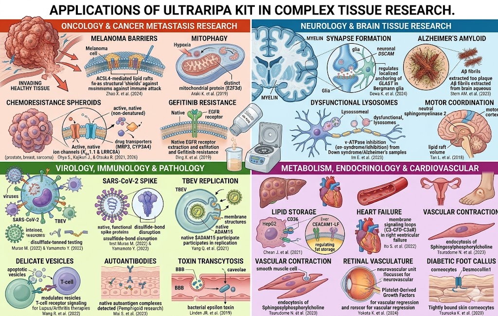
Oncology & Cancer Metastasis Research
These studies utilize the buffer to break down tight cellular structures or isolate membrane-bound signaling complexes to understand how tumors invade healthy tissue or resist chemotherapy:
-
ACSL4 & Melanoma Protective Barriers: Zhao X. et al. (2024) studied how ACSL4-mediated lipid rafts structurally prevent membrane rupture, thereby shielding melanoma cells from immunogenic cell death.
-
Mitophagy in Cancer Cells: Araki K. et al. (2019) isolated mitochondrial fractions to show how a distinct mitochondrial protein ($E2F3d$) drives hypoxia-induced mitophagy.
-
Chemoresistance Spheroid Models: A series of studies by Ohya S., Kajikuri J., and Otsuka R. (2021, 2026) used the non-denaturing qualities of the buffer on 3D tumor spheroids (sarcoma, prostate, and breast cancers) to isolate active ion channels ($K_{Ca}1.1$ and $LRRC8A$) that regulate drug transporter expressions ($MRP3$, $CYP3A4$).
-
Gefitinib Resistance in EGFR Treatment: Ding X. et al. (2019) used it to study membrane-bound epidermal growth factor receptors.
-
Mitochondrial Disruption: Liu D. (2022) looked at reversing multidrug resistance by targeting tightly bound mitochondrial membrane fractions.
-
OSCC Cell Migration: Chan NN. et al. (2023) extracted caveolae-associated proteins ($CAV1$) to study cholesterol-regulated migration in Oral Squamous Cell Carcinoma.
-
Clear Cell Renal Cell Carcinoma: Takai Y. et al. (2025) lysed cell complexes to track the interaction of $Ankrd1$ with $Talin-1$ during cell motility.
-
High-Throughput Biomarker Screening: Hayashi T. et al. (2022) extracted human osteosarcoma-specific surface antigens while keeping their structural epitopes native for phage display screening.
Neurology & Brain Tissue Research
Brain tissue is rich in myelin and complex lipid structures, making standard extraction notoriously difficult. These papers leveraged the kit to isolate functional neuro-proteins:
-
Synapse Formation & Glia: Dewa K. et al. (2024) extracted perisynaptic membrane fractions to prove that neuronal $DSCAM$ regulates the localized anchoring of $GLAST$ in Bergmann glia.
-
Alzheimer’s Disease Amyloid Fibrils: Stern AM. et al. (2023) processed aqueous extracts from human Alzheimer's disease brains to capture abundant, dense $A\beta$ fibrils from ultracentrifugal supernatants.
-
Down Syndrome & Alzheimer’s Lysosomes: Im E. et al. (2023) tracked lysosomal dysfunction caused by $v-ATPase$ inhibition using brain tissue extracts.
-
Motor Coordination: Tan L. et al. (2018) processed striatum tissue to look at neutral sphingomyelinase 2 regulation of lipid raft volume.
-
Epilepsy & Brain Activity: Tahara M. et al. (2023) analyzed brain tissue components in heterozygous $Scn1a$ knockout rat models.
Virology, Immunology & Pathology
Keeping viral spike proteins or delicate immune receptors structurally intact without denaturing them is vital for these studies:
-
SARS-CoV-2 Spike Protein Integrity: Murae M. (2022) and Yamamoto Y. (2022) isolated functional SARS-CoV-2 spike proteins to test how disulfide-bond disruptions (via glutathione or peracetic acid) impair viral functionality.
-
Tick-Borne Encephalitis Virus (TBEV): Yang Q. et al. (2021) isolated membrane structures to determine that $ADAM15$ physically participates in viral replication.
-
Lupus & Arthritis Therapies: Wang R. et al. (2022) extracted delicate apoptotic vesicles to assess how they modulate T-cell receptor signaling pathways.
-
Pemphigoid Autoantibodies: Mai S. et al. (2023) extracted a native autoantigen complex from skin tissue cells, which requires completely non-denaturing conditions to successfully detect autoantibodies.
-
Bacterial Toxin Transcytosis: Linden JR. et al. (2019) isolated caveolae fractions to track Clostridium perfringens epsilon toxin crossing the blood-brain barrier.
-
Anti-Inflammatory NF-κB Signaling: Hayakawa K. et al. (2019) utilized it for downstream evaluation of microRNA-766-3p actions.
-
CD40 Agonist Immunotherapy: Frankish J. et al. (2023) studied cell-surface antigen-presenting complex activations.
Metabolism, Endocrinology & Cardiovascular Disease
-
Lipid Storage in HepG2 Cells: Chean J. et al. (2021) isolated the fatty acid transporter $CD36$ and $CEACAM1-LF$ complexes from liver cell lines.
-
Right Ventricular Heart Failure: Ito S. et al. (2022) evaluated cardiac tissue membrane signaling loops ($C3-CFD-C3aR$).
-
Vascular Smooth Muscle Contraction: Tsurudome N. et al. (2023) tracked the endocytosis pathways of Sphingosylphosphorylcholine.
-
Retinal Vascular Regression: Yokota K. et al. (2024) analyzed neurovascular unit structural stability via platelet-derived growth factors.
-
Female Fertility Regulation: Chen LJ. et al. (2022) mapped the anchoring of $Gm364$ with $MIB2/DLL3/Notch2$ complexes to study AKT activation.
-
Pulmonary Hypertension: Kabwe JC. et al. (2022) evaluated lung tissue signaling shifts induced by a point mutation in $Bmpr2$.
-
Cell Surface Localization & Glucose: Toyoda Y. et al. (2018) extracted multipass transmembrane proteins from HeLa cell surfaces to evaluate $GRP78$ dependence.
-
Diabetic Foot Calluses: Tsuruoka K. et al. (2020) extracted Desmocollin1 tightly bound inside rigid skin corneocytes.
Related Products
Please, click below each image if you need more info.
 |
|
 |
 |
 |
 |
 |
|
 |
 |