Streptavidin, Flamma® 552
Streptavidin, Flamma® 552
Cat. No. List below
Size 1 mg
Description
Its streptavidin properties include a structure of homotetrameric protein. Its biotin binding is extremely high affinity (Kd ~ 10^-14 M). Each streptavidin molecule can bind four biotin molecules and it is a non-covalent interaction. The advantage over avidin is that it is slightly lower affinity but less non-specific binding, near-neutral pI value, and lack of carbohydrate.
Streptavidin, Flamma® 552 is suitable for a wide range of applications, including cell surface labeling, ELISA (Enzyme-Linked Immunosorbent Assay), immunohistochemistry, affinity purification, FACS (Fluorescence-Activated Cell Sorting), EMSA (Electrophoretic Mobility Shift Assay), medical diagnostics, in vitro analysis, in vivo imaging, and animal model studies.
What are the advantages?
1. Rapid binding: Quick bond formation between biotin and streptavidin
2. Stable interaction: Binding remains stable under various conditions:
a. High temperatures
b. Wide pH range
c. Presence of denaturing agents
3. Versatility: Effective detecting and analytic probe for diverse applications
4. High sensitivity: Bright fluorescence allows for detection of low-abundance targets
5. Compatibility: Suitable for in vitro, in vivo, and animal model studies
Streptavidin, Flamma® 552 works by exploiting the strong and specific interaction between streptavidin and biotin. The fluorescent label allows for easy detection and visualization of biotin-tagged molecules or structures. This conjugate can be used in both direct and indirect detection methods, depending on the specific application and experimental design.
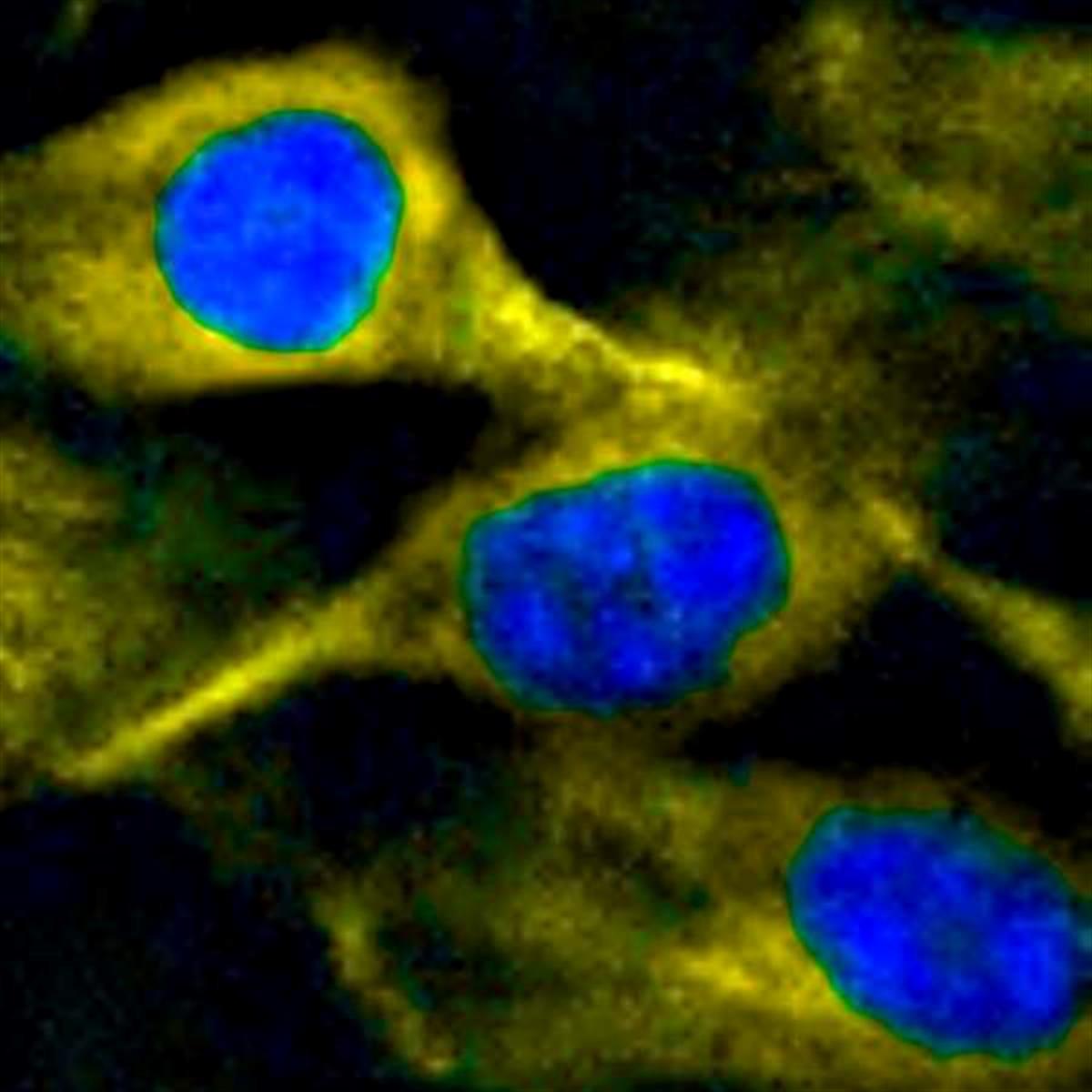
The Flamma® 552 dye is covalently attached to the streptavidin molecule, ensuring stable fluorescent labeling. This conjugate can be used in conjunction with other biotinylated molecules, such as antibodies, nucleic acids, or small molecules. The bright yellow fluorescence of Flamma® 552 makes it compatible with many common fluorescence microscopy setups and multicolor imaging experiments. Its excitation and emission properties make it suitable for use with a variety of laser lines, increasing its versatility in different imaging systems.
Streptavidin, Flamma® 552 is a highly effective and versatile tool for researchers in biochemistry, cell biology, and related fields, offering robust biotin-binding capabilities combined with bright fluorescent labeling for sensitive and specific detection in a variety of experimental contexts, including in vivo and animal model studies.
Specifications
- Fluorophore label: Flamma® 552 (bright yellow fluorescent dye)
- Reactive group: Streptavidin
- Reacting toward: Biotin
- Excitation/Emission Max.(nm): 550/565
- Excitation: Can be excited using 532, 543, 546, or 555 nm laser lines
- Emission: Bright yellow fluorescence
- Storage conditions: 4 ℃, protect from light
Table 1. List of fluorescent dye conjugates of Streptavidin
| Quick link (Cat.#) | Series | Ex * (nm) | Em* (nm) |
| RFP0716 | Streptavidin, Flamma® 496 | 496 | 516 |
| RFP0705 | Streptavidin, Flamma® 552 | 550 | 565 |
| RFP0711 | Streptavidin, Flamma® 648 | 648 | 663 |
| RFP0712 | Streptavidin, Flamma® 675 | 675 | 691 |
| RFP0713 | Streptavidin, Flamma® 749 | 749 | 774 |
| RFP0714 | Streptavidin, Flamma® 774 | 774 | 806 |
Table 2. List of fluorescent dye conjugates of Biotin
| Quick link (Cat.#) | Series | Ex * (nm) | Em* (nm) | Molar mass (g/mol) |
| RFP0616 | Biotin, Flamma® 496 | 496 | 516 | 654.64 |
| RFP0605 | Biotin, Flamma® 552 | 550 | 565 | 913.18 |
| RFP0611 | Biotin, Flamma® 648 | 648 | 663 | 927.20 |
| RFP0612 | Biotin, Flamma® 675 | 675 | 691 | 1185.43 |
| RFP0613 | Biotin, Flamma® 749 | 749 | 774 | 951.22 |
| RFP0614 | Biotin, Flamma® 774 | 774 | 806 | 1169.25 |
Background
Streptavidin is smaller (53 KDa) and has a little lower affinity than avidin yet displays less non-specific binding due to near-neutral pI value and lack of carbohydrates. Biotin, a 244 dalton vitamin found in all living cells, binds with high affinity to avidin and streptavidin. In biotechnology, biotin is conjugated to antibodies, enzymes, reporter to form the tetravalent binding nature of biotin-avidin/streptavidin complex. Biotinavidin/streptavidin binding has high affinity, which has been utilized in diverse applications such as ELISA, immunohistochemistry, cell surface labeling, affinity purification, FACS, EMSA, etc. The bond formation between biotin and avidin/streptavidin is very rapid, and once formed, is stable at high temperature and in a wide range of pH, organic solvents and denaturing agents. The system is a simple yet elegant and can be incorporated into virtually every immunoassay where an antibody is conjugated with biotin and then detected with avidin or streptavidin conjugated to various commercially available fluorophores and reporters. BioActs offers a variety of fluorescent dye conjugated streptavidin and biotin as effective detecting and analytic probes for diverse applications in biochemical and biological research fields.
♦ Conjugated with a wide range of fluorescent dyes
♦ Can be utilized in a variety of applications.
♦ Bright and photostable fluorescence
♦ High water solubility
Fluorescent Streptavidin & Biotin
Avidin and streptavidin are both tetrameric proteins composed of four identical subunits, each bind four biotins (vitamin H) per molecule with high binding affinity and specificity (Kd ~ 1015 M for avidin and ~ 1014 M for streptavidin). Although the primary sequence homogeneity of both proteins are 30%, their tertiary and quaternary structure are almost identical, and anti-avidin and anti-streptavidin antibodies are not immunologically cross reactive. Avidin, a 67 KDa glycoprotein with an isoelectric point of about 10.5, has the higher affinity than streptavidin, however it also displays more nonspecific binding and aggregation due to its oligosaccharide component (mannose and N-acetylglucosamine) and positive charge. Streptavidin is smaller (53 KDa) and has a little lower affinity than avidin yet displays less non-specific binding due to near-neutral pI value and lack of carbohydrates. Biotin, a 244 dalton vitamin found in all living cells, binds with high affinity to avidin and streptavidin. In biotechnology, biotin is conjugated to antibodies, enzymes, reporter to form the tetravalent binding nature of biotin-avidin/streptavidin complex. The valeric acid side of biotin can be incorporated with various functional groups, reporters and fluorophores that can be utilized in a wide range of biological structures and processes.
Biotin-avidin/streptavidin binding has high affinity, which has been utilized in diverse applications such as ELISA, immunohistochemistry, cell surface labeling, affinity purification, FACS, EMSA, etc. The bond formation between biotin and avidin/streptavidin is very rapid, and once formed, is stable at high temperature and in a wide range of pH, organic solvents and denaturing agents. The system is a simple yet elegant and can be incorporated into virtually every immunoassay where an antibody is conjugated with biotin and then detected with avidin or streptavidin conjugated to various commercially available fluorophores and reporters. These features of biotin and avidin/streptavidin are useful for purifying or detecting proteins conjugated to either component of the interaction. Although biotin-avidin/streptavidin system is simple and easy to use, it also has some limitations: biotinylated compounds might non-selectively bind to any biotin-binding protein, endogenous biotin can cause background noise, and harsh conditions are needed to break their interaction that might limit its application. BioActs offers a variety of fluorescent dye conjugated avidin, streptavidin and biotin as effective detecting and analytic probes for diverse applications in biochemical and biological research fields.
Preparation of fluorescence labeled Streptavidin/Biotin solutions
To dissolve dye-labeled compound powder in 0.5–1.0 mL of PBS or other suitable buffer.
The dye-conjugates are stable for at least one years when stored as directed.
For longer storage, divide solutions into aliquots and freeze at <–20°C.
Avoid from light, repeated freezing and thawing of solutions.
Labeling with conjugates of Streptavidin
Streptavidin conjugates are used as secondary detection reagents in many biotechnical applications.
These reagents can also be employed to bind biotin and its derivatives.
♦ Direct Streptavidin labeling procedure: Biotin-labeled primary probes such as antibodies, nucleic acids or lectins are conjugated to tissues, cell surfaces or other biomolecules. Excess protein is removed by washing, and detection is facilitated by fluorescent avidin/streptavidin.
♦ Indirect Streptavidin labeling procedure: Biotin-labeled antibodies or oligonucleotides are conjugated to tissues, cell surfaces or other biomolecules. This preparation is then treated with unlabeled streptavidin, and excess reagents are removed by washing. Detection is realized by treating of fluorescent biotin derivatives.
Alternatively, an unlabeled primary antibody is attached to a biomolecular target, which would be bound by the biotinlabeled secondary antibody. The complex is detected by the direct or indirect procedures described above.
♦ Centrifuge protein conjugate solutions briefly before using, and only the supernatant should be used for the experiment in order to eliminate any aggregates, thereby reducing the background signal.
Staining protocols may vary depending on the experimental condition, thus determine appropriate dilution for conjugates
empirically.
Citation & Reference
1. Ting-Wei Wu. Fluorescent Probe Encapsulated in Avidin Protein to Eliminate Nonspecific Fluorescence and Increase Detection Sensitivity in Blood Serum. Anal Chem 88.16 (2016): 7873-7.
2. Qian Sun. Discrimination between streptavidin and avidin with fluorescent affinity-based probes. Analyst 140 (2015): 4648-4653.
3. Yung-Peng Wu. Target-activated streptavidin–biotin controlled binding probe. Chem. Sci 9 (2018): 770-776.
4. Akshay Jain. The principles and applications of avidin-based nanoparticles in drug delivery and diagnosis. Journal of Controlled Release 245.10 (2017): 27-40.
5. MEIR WILCHEK. Introduction to Avidin-Biotin Technology. Methods Enzymol 184 (1990): 5-13.
6. JEANNE BENTLEY LAWRENCE. Interphase and Metaphase Resolution of Different Distances Within the Human Dystrophin Gene. Science New Series 249.4971 (1990): 928-932.