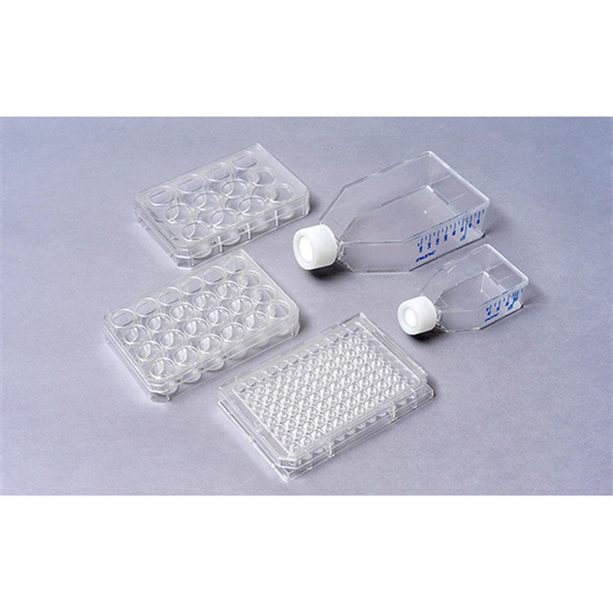

HydroCell, T75 Flask
Product#: FNK-CSF075
$556.29
Availability:
Ships in 1-2 Weeks
HydroCell (Flask)
Cat No: List below
Product ID and Size
| Quick Link | Products | Cat. No. | Packing Unit (pcs) | Size (mm) | Surface Area | Capacity per well | Link |
| HydroCell (Flask) | T25 Flask | FNK-CSF025 | 12 | 81(W)×125(L) | 25 cm2 | 70 mL |  |
| T75 Flask | FNK-CS2015 | 5 | 40(D)×12(H) | 8.8 cm2 | 3 mL | |
|
Description
Hydrocell is a low cell binding cultureware. On its surface, super hydrophilic polymer was immobilized at nano-thickness.
This hydrophilic character helps the formation of embryoid bodies of ES/iPS cell or Spheroid in culture.
Compared to other brands' low cell binding cultureware, HydroCell shows lower cell binding.
Features
- Super hydrophilic polymer is immobilized to the surface of the cultureware at nano-thickness.
- Lowest attachment rate among similar competitive brands.
- Three formats : Microplates, Dishes and Flask types.
Best for...
- Formation of embryoid bodies of ES cells in culture
- Formation of anchorage-independent colonies
- Differentiation such as cartilage (spheroid formation)
- Culture & storage of macrophage/immune cells
- Screening of anti-cancer drugs (replacement for soft agar assays)
| HydroCell Surface Designed to Prevent Cell Attachment |
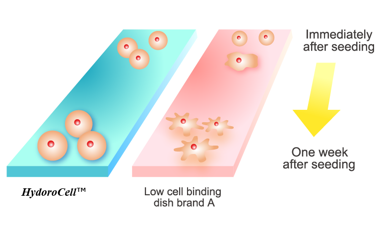 |
Image of HydroCell surface Super hydrophilic polymer is immobilized to the surface of the cultureware at nano-thickness. Suitable for
|
| Comparison to Other Products |
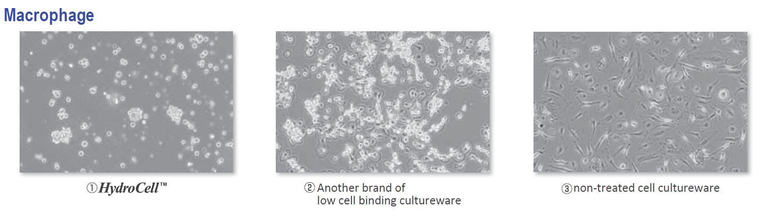 |
| Macrophages from mouse peritoneum in medium with serum were incubated for 3 days in (1) cultureware with HydroCell (2) another brand of low cell binding cultureware, and (3) non-treated cell cultureware. Only the HydroCell allowed uniform suspension culture (more than 99% of the cells remained in suspension) and simple cell dissociation and harvesting by pipetting. |
| Lowest Cell Attachment Rate among Competitive Brands |
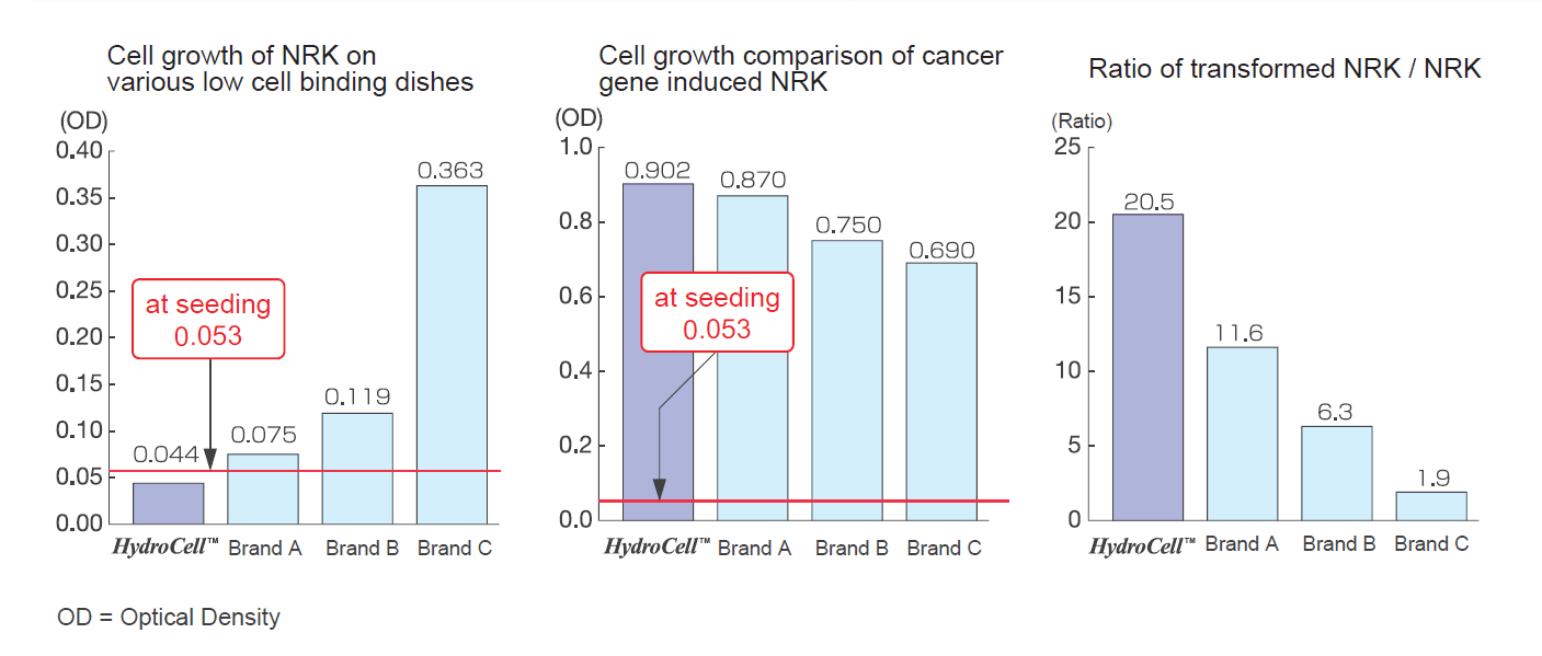 |
| Nomal Rat Kidney (NRK) cells in medium with serum were seeded in cultureware with HydroCell or in a low cell binding product of brand A, B or C. Degree of cell adhesion was determined using a TetraColor ONE assay system (Seikagaku Corp.; Tokyo, Japan) at seeding and after 5 days of incubation. |
| Competitive evaluation of HydroCell™ for embryoid body formation of ES Cells (human) |
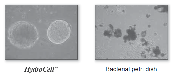 |
Left : Uniform suspension culture with HydroCell Right : Cell adhesion on bacterial petri dish *Cultured in medium with 20% FBS |
| Example of a Differention Induction |
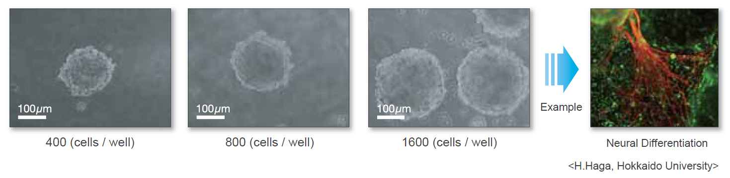 |
| Murine ES cells (B6G-2) were incubated for 2 days in HydroCell 96MW Photos taken on slide glass |
| Application for Anti-cancer Drugs Screening |
Noise produced by growth of non-cancer cells mixed with cancer cells can be inhibited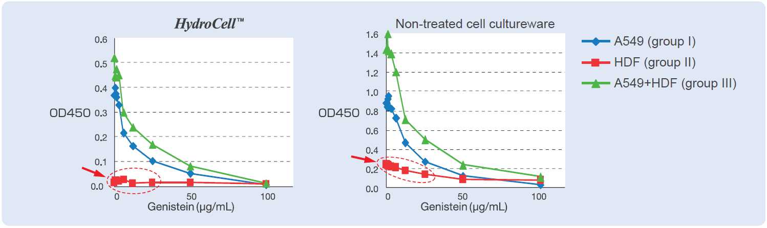 |
| Protocol : 1. The following cells were seeded in HydroCell 96MW and non-treated cell cultureware group I : A-549 (2.0 X 10³ cells)
group II : Human Dermal Fibroblasts (HDF, 2.0 X 10³ cells)
group III : A-549 (2.0 X 10³ cells) + Human Dermal Fibroblasts (HDF, 2.0 X 10³ cells)
2. Genistein, concentration range 0 - 100ug / mL, was added to each well and incubated for 3 days.3. 10uL of live cell counting reagent was added to each well, and the absorbance OD450 was measured after 4 hours. |
| Stem Cell Sphere Formation |
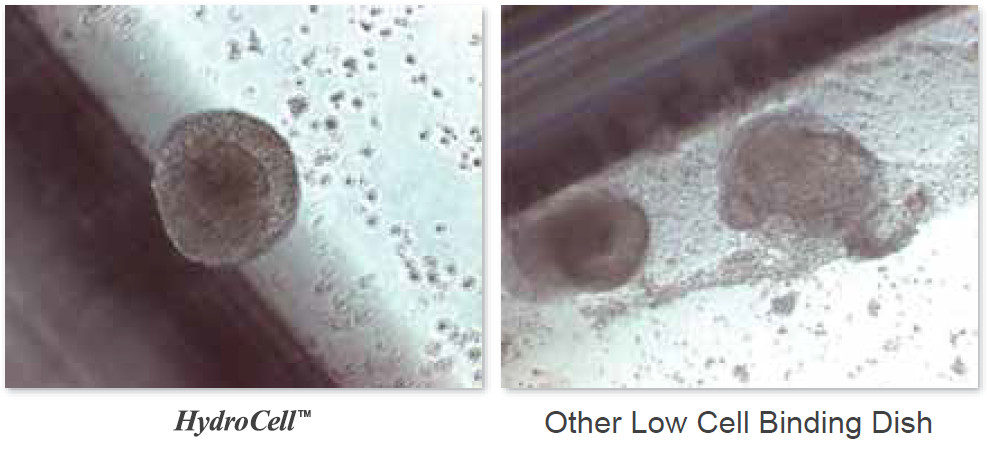 |
(A) hiPS Cells Sphere Formation iPS spheres are formed on HydroCell (left photo) compared to the adherence to the edge of other brand dish (right photo) <A. Umezawa, National Center for Child Health and Development> |
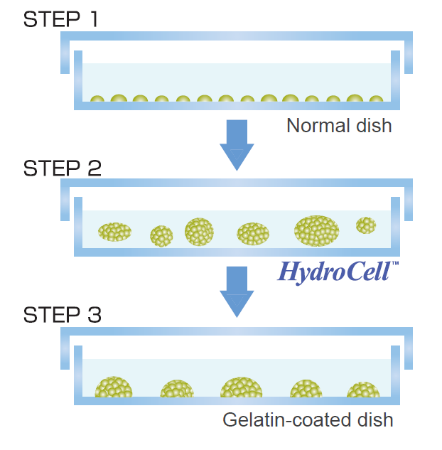 |
(B) hES Cells Sphere Formation Method A short time floating culture is extremely useful as a way to sphere formation in the induction of differentiation on feeder-free conditions. Step1: hES cells maintained without feeder cells Step2: Sphere formation on HydroCell Step3: Adherent culture of spheres on a gelatin-coated dish <Kumiko Saeki, International Medical Center of Japan, Tokyo, Japan> |
This item is oversize shipping by Volume weight. ( 21x19x14, inch)