.jpg "/Content/Upload/Product/Human_ELISA_kits_Diagnocine_1200_1200(101).jpg")
Human mammary carcinoma marker CA15-3 (CA15-3) ELISA kit
The Ultimate 1-Stop ELISA Marketplace:
60,000+ ELISA kits Available to Serve Scientists from All Fields of Life Science
Always Powered by Diagnocine for Precision Research.
Most comprehensive buffers, reagents and detection agents are provided with each kit to optimize data output.
All-inclusive format covers everything from sample preparation to signal detection. Every step can be customized and optimized for your precision research.
DiagnoCine Precision buffers and reagents are Made in USA ISO 13485 Certified
Why waste time searching for your ELISA kit when you can get everything in one place? Diagnocine can provide you with all the ELISA needs you have (customized ELISAs also available) on the planet through a single, streamlined ordering process.
* all types of species
* HRP and AP detection
* Low target signal enhancement
* Inquire about a trial version or a customized version
4 core reagents below are included with every order, plus 1 of the 2 detection-specific reagents (matched automatically to your kit type) — 5 components, every time.
+ the 5th component, matched to your kit's detection method:
If you have any questions or need additional information, please call us at 551-245-1033 or email us at support@diagnocine.com. Our team will be happy to assist you.
Universal Compatibility
All buffers and detection reagents work seamlessly across target proteins, bridging the gap in assay consistency.
1-Stop Convenience
Consolidate your lab's purchasing. Source all ELISA needs and experience optimal buffers and reagents in a single shipment.
Enhanced Precision
Designed by scientists for scientists, all components are engineered to deliver sharper results and an optical signal.
Human mammary carcinoma marker CA15-3 (CA15-3) ELISA kit
Sandwich quantitative immunoassay for Human mammary carcinoma marker CA15-3 (CA15-3) ELISA kit in human serum available in multiple catalog sizes:
Note: Please send inquiries regarding Trial 24T orders to support@diagnocine.com.
| Uniprot No. | P15941 |
| Species | Homo sapiens (Human) |
| Sample types | serum |
| Detection range | 15 U/mL - 250 U/mL |
| Sensitivity | 3.8 U/mL |
| Assay time | 1-5h |
| Sample loading volume | 50-100μL |
| Detection wavelength | 450 nm |
| Assay principle | Sandwich (Quantitative) |
| Data analysis | Standard curve + Curve Expert software |
| Research area | Cancer |
| Storage condition | 2-8°C (see protocol for full details) |
| Shipping condition | 4 °C |
| Shelf life | 6 months |
10 business days
Processing + 3-5 days shipping
In Stock : USA
Worldwide shipping available
Antibody capture
450 nm detection
Standard curve
Quantification
In this sandwich ELISA, Human mammary carcinoma marker CA15-3 (CA15-3) ELISA kit in the sample is captured between a pre-coated capture antibody and a detection antibody. Signal intensity is proportional to analyte concentration. Quantification uses a standard curve fitted with Curve Expert software, covering 15 U/mL - 250 U/mL with a minimum detectable dose of 3.8 U/mL.
This Human CA15-3 ELISA Kit was designed for the quantitative measurement of Human CA15-3 protein in serum. It is a Sandwich ELISA kit, its detection range is 15 U/mL - 250 U/mL and the sensitivity is 3.8 U/mL.
- findings suggest that these pulmonary markers could be useful to assess CAP severity and, especially YKL-40 and CCL18 by helping predict CAP caused by atypical pathogens PMID: 29324810
- Quercetin suppressed breast cancer stem cell proliferation, self-renewal, and invasiveness. It also lowered the expression levels of proteins related to tumorigenesis and cancer progression, such as aldehyde dehydrogenase 1A1, C-X-C chemokine receptor type 4, mucin 1, and epithelial cell adhesion molecules. PMID: 29353288
- We have analysed the tumour-associated carbohydrate antigens sialyl-Lewis x (SLe(x)) and sialyl-Tn (STn) on MUC1 and MUC5AC in Pancreatic adenocarcinoma (PDAC)tissues. immunoprecipitation of MUC5AC from positive PDAC tissues and subsequent SLe(x) immunodetection confirmed the presence of SLe(x) on MUC5AC. Altogether, MUC5AC-SLe(x) glycoform is present in PDAC and can be regarded as potential biomarker. PMID: 29408556
- High MUC1 expression is associated with breast cancer metastasis. PMID: 29433529
- In the in vitro tests, JFD-WS effectively inhibited HUVEC proliferation, migration, tube formation and VEGFR2 phosphorylation. Additionally, JFD-WS inhibited the formation of blood vessels in chick chorioallantoic membrane. While inhibiting the xenograft tumor growth in experimental mice, JFD-WS decreased the plasma MUC1 levels PMID: 29436685
- KL-6 is an accurate biomarker for the diagnosis of interstitial lung disease in systemic sclerosis. PMID: 29455320
- we wanted to explore whether STAT3 can be related to lymph node micrometastasis of non-small cell lung cancer (NSCLC). To address this question, we evaluated the expression of MUC1 mRNA in the lymph node samples of NSCLC to determine micrometastasis. Then, we evaluated what role STAT3 overexpression plays in lymph node micrometastasis of NSCLC. PMID: 29575778
- MUC1 was a potential molecular target may help explain the role of lincRNA-ROR/miR-145 for invasion and metastasis in Triple-negative breast cancer cell lines. PMID: 29673594
- The glycosylation level of CA153 was found to increase with increasing breast cancer stage in the sandwich assay. The assay system appeared to efficiently discriminate breast cancer stage I (sensitivity: 63%, specificity: 69%), IIA (sensitivity: 77%, specificity: 75%), IIB (sensitivity: 69%, specificity: 86%) and III (sensitivity: 80%, specificity: 65%) from benign breast disease. PMID: 29749490
- Results show MUC1 expression highly expressed at mRNA and protein levels in esophageal squamous cell carcinoma (ESCC). MUC1 expression correlated with tumor invasion, lymph node metastasis, and TNM staging. PMID: 29798942
- Decreased expression of MUC1 is an independent marker for endometrial receptivity in recurrent implantation failure. PMID: 29929546
- The expression profile of studied Mucins MUC16 and MUC1 and truncated O-glycans was not associated with the site of origin of ovarian cancer (OVCA) cell lines PMID: 30011875
- High MUC1 expression is associated with cervical cancer. PMID: 30062487
Intra-assay Precision (Precision within an assay): CV%<15% | ||||||
Three samples of known concentration were tested twenty times on one plate to assess. | ||||||
Inter-assay Precision (Precision between assays): CV%<20% | ||||||
Three samples of known concentration were tested in twenty assays to assess. | ||||||
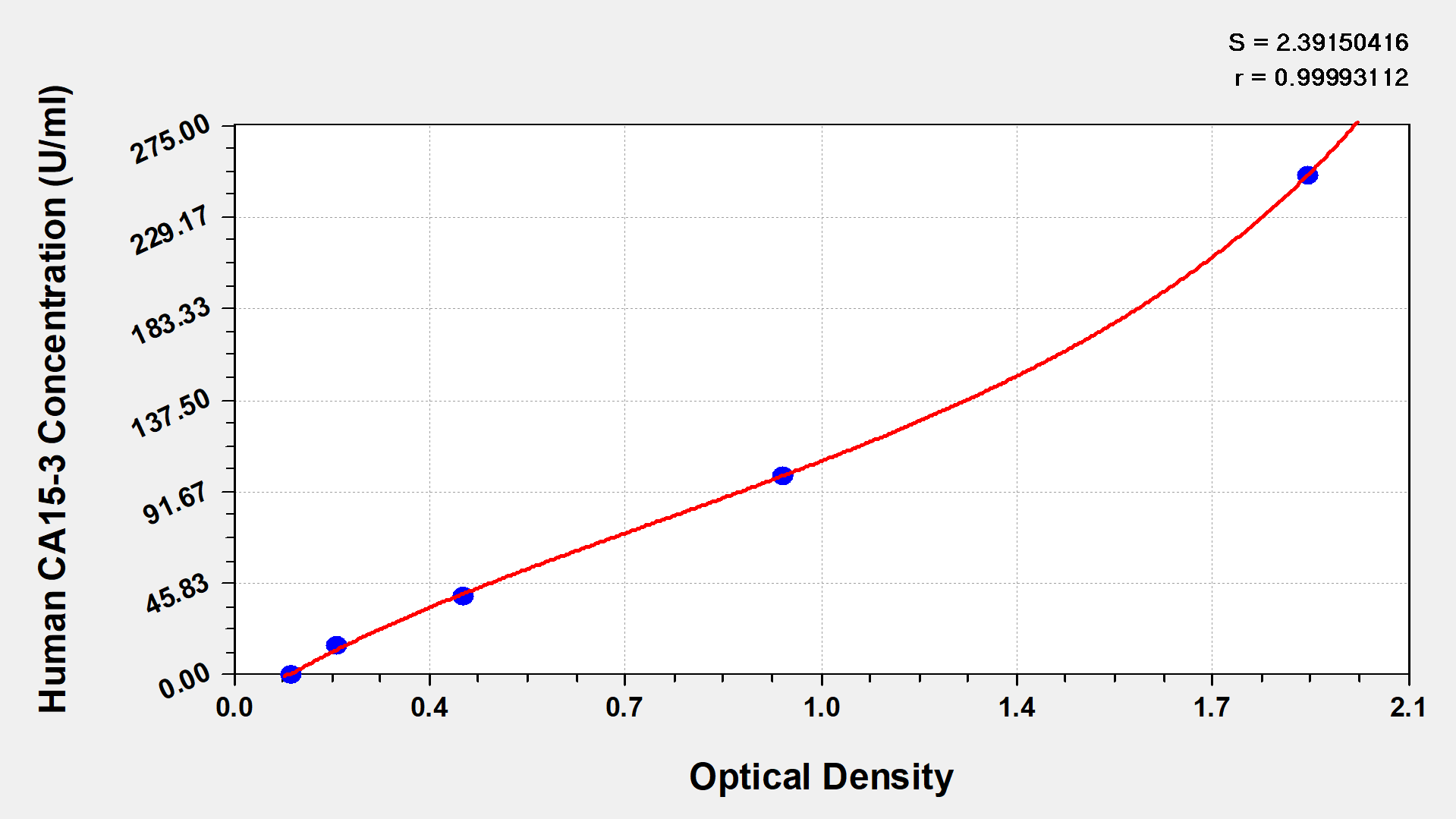
These standard curves are provided for demonstration only. A standard curve should be generated for each set of samples assayed. | |||||||
| |||||||
U/ml | OD1 | OD2 | Average |
| |||
0 | 0.109 | 0.114 | 0.112 |
| |||
15 | 0.187 | 0.196 | 0.192 |
| |||
40 | 0.425 | 0.401 | 0.413 |
| |||
100 | 0.964 | 0.975 | 0.970 |
| |||
250 | 1.937 | 1.836 | 1.887 | ||||
To assess the linearity of the assay, samples were spiked with high concentrations of human CA15-3 in various matrices and diluted with the Sample Diluent to produce samples with values within the dynamic range of the assay. | ||||||
| Sample | Serum(n=4) | ||||
1:1 | Average % | 90 | ||||
Range % | 86-99 | |||||
1:2 | Average % | 97 | ||||
Range % | 91-105 | |||||
1:4 | Average % | 95 | ||||
Range % | 89-100 | |||||
1:8 | Average % | 92 | ||||
Range % | 88-98 | |||||
The recovery of human CA15-3 spiked to levels throughout the range of the assay in various matrices was evaluated. Samples were diluted prior to assay as directed in the Sample Preparation section. | ||||||
Sample Type | Average % Recovery | Range | ||||
Serum (n=5) | 97 | 93-99 | ||||
EDTA plasma (n=4) | 96 | 90-101 | ||||
- A micro ELISA plate --- The 96-well plate has been pre-coated with an anti-human CA15-3 antibody.
- Five vials standard (0.5 ml/bottle)--- Dilute a bottle of the standard at dilution series, read the OD values, and then draw a standard curve.
- One vial HRP-conjugated CA15-3 antibody (6 ml/bottle) --- Bind to the CEA in the samples or standards and react with the substrate to make the solution chromogenic.
- One vial Sample Diluent (11ml/bottle) ---Dilute the sample solution.
- One vial Wash Buffer (20x concentrate) (15 ml/bottle) --- Wash away unbound or free substances.
- One vial Substrate A (7ml/bottle) --- Mix with substrate B and then interact with TMB to elicit a chromogenic reaction.
- One vial Substrate B (7ml/bottle) --- Mix with substrate A and interact with TMB to elicit a chromogenic reaction.
- One vial Stop Solution (7ml/bottle) --- Stop the color reaction. The solution color immediately turns from blue to yellow.
- Four Adhesive Strips (For 96 wells) --- Cover the microplate when incubation.
- An instruction manual
- A microplate reader capable of measuring absorbance at 450 nm, with the correction wavelength set at603 nm or 630 nm.
- An incubator can provide stable incubation conditions up to 37°C±5°C.
- Centrifuge
- Vortex
- Squirt bottle, manifold dispenser, or automated microplate washer
- Absorbent paper for blotting the microtiter plate
- 50-300ul multi-channel micropipette
- Pipette tips
- Single-channel micropipette with different ranges
- 100ml and 500ml graduated cylinders
- Deionized or distilled water
- Timer
- Test tubes for dilution