.jpg "/Content/Upload/Product/Human_ELISA_kits_Diagnocine_1200_1200(420).jpg")
Human Seprase(FAP) ELISA kit
The Ultimate 1-Stop ELISA Marketplace:
60,000+ ELISA kits Available to Serve Scientists from All Fields of Life Science
Always Powered by Diagnocine for Precision Research.
Most comprehensive buffers, reagents and detection agents are provided with each kit to optimize data output.
All-inclusive format covers everything from sample preparation to signal detection. Every step can be customized and optimized for your precision research.
DiagnoCine Precision buffers and reagents are Made in USA ISO 13485 Certified
Why waste time searching for your ELISA kit when you can get everything in one place? Diagnocine can provide you with all the ELISA needs you have (customized ELISAs also available) on the planet through a single, streamlined ordering process.
* all types of species
* HRP and AP detection
* Low target signal enhancement
* Inquire about a trial version or a customized version
4 core reagents below are included with every order, plus 1 of the 2 detection-specific reagents (matched automatically to your kit type) — 5 components, every time.
+ the 5th component, matched to your kit's detection method:
If you have any questions or need additional information, please call us at 551-245-1033 or email us at support@diagnocine.com. Our team will be happy to assist you.
Universal Compatibility
All buffers and detection reagents work seamlessly across target proteins, bridging the gap in assay consistency.
1-Stop Convenience
Consolidate your lab's purchasing. Source all ELISA needs and experience optimal buffers and reagents in a single shipment.
Enhanced Precision
Designed by scientists for scientists, all components are engineered to deliver sharper results and an optical signal.
Human Seprase(FAP) ELISA kit
Sandwich quantitative immunoassay for Human Seprase(FAP) ELISA kit in human serum, plasma,tissue homogenates available in multiple catalog sizes:
Note: Please send inquiries regarding Trial 24T orders to support@diagnocine.com.
| Uniprot No. | Q12884 |
| Species | Homo sapiens (Human) |
| Sample types | serum, plasma,tissue homogenates |
| Detection range | 0.312 ng/mL-20 ng/mL |
| Sensitivity | 0.078 ng/mL |
| Assay time | 1-5h |
| Sample loading volume | 50-100μL |
| Detection wavelength | 450 nm |
| Assay principle | Sandwich (Quantitative) |
| Data analysis | Standard curve + Curve Expert software |
| Research area | Cancer |
| Storage condition | 2-8°C (see protocol for full details) |
| Shipping condition | 4 °C |
| Shelf life | 6 months |
10 business days
Processing + 3-5 days shipping
In Stock : USA
Worldwide shipping available
Antibody capture
450 nm detection
Standard curve
Quantification
In this sandwich ELISA, Human Seprase(FAP) ELISA kit in the sample is captured between a pre-coated capture antibody and a detection antibody. Signal intensity is proportional to analyte concentration. Quantification uses a standard curve fitted with Curve Expert software, covering 0.312 ng/mL-20 ng/mL with a minimum detectable dose of 0.078 ng/mL.
This Human FAP ELISA Kit was designed for the quantitative measurement of Human FAP protein in serum, plasma,tissue homogenates. It is a Sandwich ELISA kit, its detection range is 0.312ng/mL-20 ng/mL and the sensitivity is 0.078 ng/mL.
- the circulating protease, fibroblast activation protein, is the proteolytic enzyme responsible for hFGF21 inactivation. PMID: 26635356
- DPP4 activity and/or structure homologue (DASH) proteins are involved in many pathophysiological processes and have therefore been proposed for potential biomarkers or even drug targets in various cancers (DPP4 and FAP). (Review) PMID: 26671446
- Increased FAPalpha expression is associated with thyroid papillary carcinoma. PMID: 26715280
- This study identified fibroblast activation protein (FAP) as the enzyme that cleaves and inactivates human FGF21. PMID: 26797127
- FAP selectively cleaves type I collagen resulting in increased macrophage adhesion. PMID: 26934296
- Human FGF-21 Is a Substrate of Fibroblast Activation Protein. PMID: 26962859
- Data suggest that a DNA vaccine targeting human fibroblast activation protein alpha (FAPalpha) may be an attractive and effective cancer immunotherapy strategy. PMID: 27020681
- Fibroblast activation protein (P=.00117) was stronger than grade and stage in predicting clinical aggressiveness in clear cell renal cell carcinoma. PMID: 27063470
- have identified fibroblast activation protein (FAP) as the endopeptidase responsible for this site-specific cleavage of human FGF21 (hFGF21), and propose that inhibition of FAP may be a therapeutic strategy to increase endogenous levels of active FGF21. PMID: 27118870
- Mutations to predicted TM interfacial residues (G10L, S14L, and A18L) comprising a small-X3-small motif reduced FAP TM-CYTO dimerization relative to wild type. Predicted off-interface residues showed no significant change from wild type. The interfacial TM residue G10L decreased FAP endopeptidase activity more than 25%, and reduced cell-surface versus intracellular expression relative to interfacial S14L and A18L. PMID: 27155568
- High FAPalpha expression is associated with glioblastoma. PMID: 27492457
- The level of FAP expression in NGP-127, SJCRH30, and SJSA-1 lines as well as in cancer-associated fibroblasts of patients was comparable, which makes these cell lines a possible model for studying FAP PMID: 27817025
- Expression of FAPalpha in stroma was associated with distant metastasis of breast phyllodes tumor. PMID: 27881889
- This evidence highly suggested that FAP is a potential prognosticator of GC patients and a target for synergizing with other treatments, especially immune checkpoint blockades in GC. PMID: 27983931
- FAP expression is significantly upregulated in human masticatory mucosa during wound healing PMID: 28005267
- expression of FAP in primary tumors and in their metastases was associated both with synchronous metastases and also with metastases to the lymph nodes PMID: 28033421
- These results indicated that the low plasma FAPalpha level might due to the systemic reaction to the presence of tumor and circulating FAPalpha level might be a potential indicator for diagnosing ESCC. PMID: 28415791
- Several isoforms of DPP-IV and FAP are present in glioblastoma tissue. The absence of alkaline isoforms of both enzymes in glioma cell lines however suggests that isoforms from other, most likely stromal, cell types contribute to the overall pattern seen in glioblastoma tissues. PMID: 28452380
- The predictors for FAP occurrence among desmoid tumor patients are large tumor size, intra-abdominal location, multiple tumors, and patient's young age. PMID: 28570749
- Circulating FAP activity and antigen levels correlate strongly when measured in liver disease and coronary heart disease. PMID: 28582421
- proCOL11A1, fibroblast-activated protein, secreted protein acidic and rich in cysteine, and periostin expression was significantly increased in the intratumoral stroma of pancreatic ductal adenocarcinomas compared to paired non-neoplastic pancreata PMID: 29025374
- Mounting evidence supported that miR-30a-5p directly targetted FAP and suppressed its expression in oral cavity cancer cells (OSCCs). By suppressing FAP expression, miR-30a-5p significantly inhibited cell propagation, migration, and invasion. Therefore, miR-30a-5p might be a new therapeutic target for oral cancer treatment. PMID: 29026005
- These results revealed that FAPalpha promoted the growth, adhesion and migration of lung squamous cell carcinoma cells. In addition, FAPalpha regulated lung cancer cell function, potentially via the PI3K and SHH pathways. Further investigations are required to examine the role of FAPalpha in lung AC cells. PMID: 29115573
- In this study, authors confirm that FAPalpha can promote the generation of Tregs and TAMs, which suggests that FAPalpha plays a immunosuppressive role in the tumor microenvironment PMID: 29273462
- FAP is virtually absent in normal tissues, but it is present in the embryonic and tumor tissues, which makes it a selective and versatile model. In this work, basic approaches to affecting the CAF using FAP as a target were discussed. PMID: 30383930
Intra-assay Precision (Precision within an assay): CV%<8% | ||||||
Three samples of known concentration were tested twenty times on one plate to assess. | ||||||
Inter-assay Precision (Precision between assays): CV%<10% | ||||||
Three samples of known concentration were tested in twenty assays to assess. | ||||||
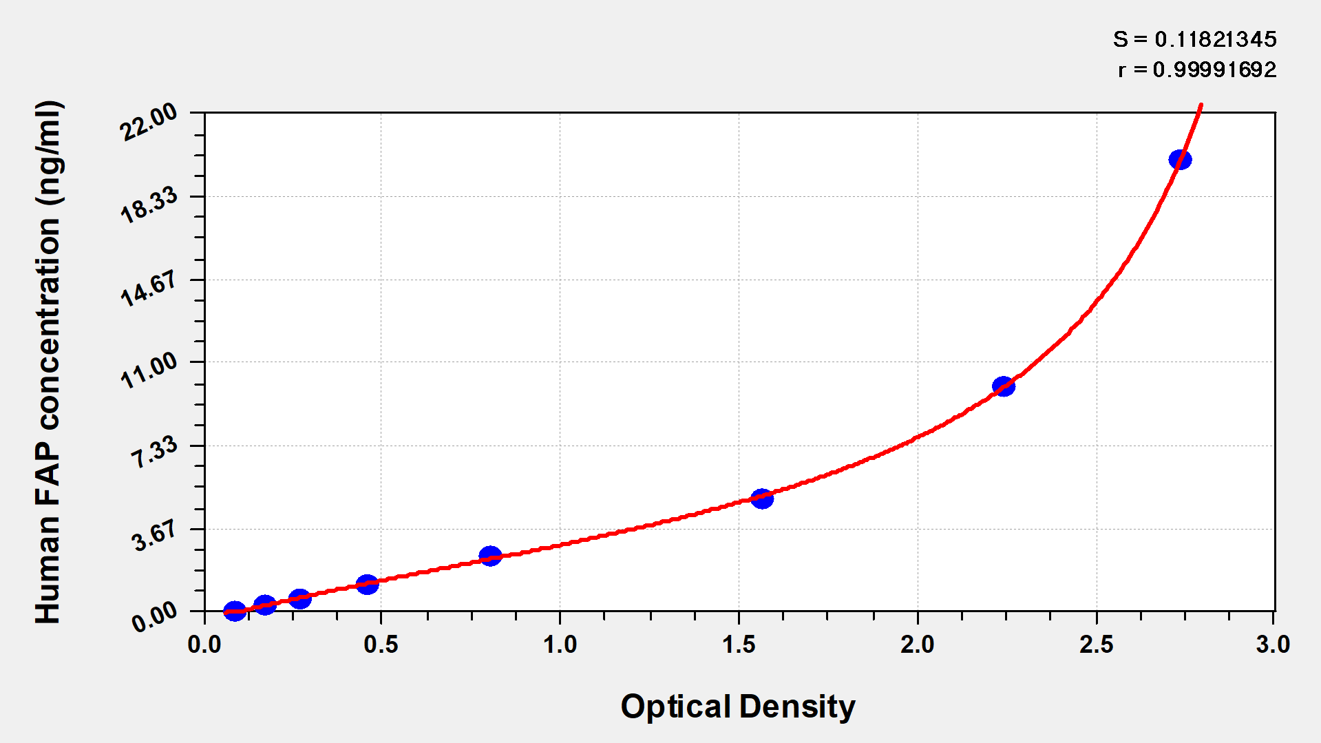
These standard curves are provided for demonstration only. A standard curve should be generated for each set of samples assayed. | |||||||
| |||||||
ng/ml | OD1 | OD2 | Average | Corrected | |||
20 | 2.864 | 2.655 | 2.760 | 2.665 | |||
10 | 2.308 | 2.214 | 2.261 | 2.166 | |||
5 | 1.637 | 1.533 | 1.585 | 1.490 | |||
2.5 | 0.827 | 0.805 | 0.816 | 0.721 | |||
1.25 | 0.476 | 0.467 | 0.472 | 0.377 | |||
0.625 | 0.289 | 0.272 | 0.281 | 0.186 | |||
0.312 | 0.184 | 0.178 | 0.181 | 0.086 | |||
0 | 0.096 | 0.094 | 0.095 |
| |||
To assess the linearity of the assay, samples were spiked with high concentrations of Human FAP in various matrices and diluted with the Sample Diluent to produce samples with values within the dynamic range of the assay. | ||||||
| Sample | Serum(n=4) | ||||
1:2 | Average % | 99 | ||||
Range % | 95-103 | |||||
1:4 | Average % | 87 | ||||
Range % | 83-93 | |||||
1:8 | Average % | 95 | ||||
Range % | 89-99 | |||||
1:16 | Average % | 88 | ||||
Range % | 82-94 | |||||
The recovery of Human FAP spiked to levels throughout the range of the assay in various matrices was evaluated. Samples were diluted prior to assay as directed in the Sample Preparation section. | ||||||
Sample Type | Average % Recovery | Range | ||||
Serum (n=5) | 89 | 85-93 | ||||
EDTA plasma (n=4) | 94 | 88-98 | ||||