HBsAg M-protein(Recombinant)
Product#: FNK-BCL-AGM-01
$1,473.78
Availability:
Ships in 1-2 Weeks
HBsAg,Pre-S1 peptide(gtA)(Recombinant, Tagged)
Cat. No.: FNK-BCL-AGS1A-21
Size: 100μg
Storage: -20℃ (3 years)
Introduction:
HBsAg (also called Bionanocapsule) is the surface antigen of Hepatitis B virus (HBV). HBsAg is known to be present in blood if HBV infected patients, and used as a important diagnostic maker of HBV infection. HBsAg forms a small lipid particle(diameter 50 to 60nm)in which many antigen protein are present. Each antigen protein has three domains, called S, Pre-S1 and Pre-S2. L-type antigen contains all the three domains, M-type antigen contains S- and Pre-S2, and S-type antigen consisted of only S-domains. During the process of HBV infection, Pre-S1 domain plays a key role for host (hepatic) cell recognition, and Pre-S2 domain is essential for cell penetration. In infected patients, hepatic cells produces a lot of non-infectious S-type HBsAg (HBV mimetics without genome). The detection of serum Pre-S2 and Pre-S1 antigen is, therefore, very important to reveal the mechanism of HBV infection. Beacle has over a decade of R&D history on HBsAg and related materials, and developed the production process of various HBsAgs.
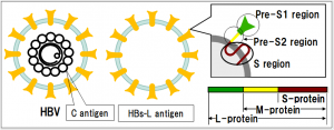
Hepatitis B virus (HBV) expresses three types of surface antigens, i.e. S-, M-, and L-protein. L-protein is composed of S-, Pre-S2, and Pre-S1 region. The deletion of Pre-S1 region forms M-protein and further deletion of Pre-S2 region results in S-protein. Most of commercially available HBsAg is composed of either S-protein alone or a mixture of Sand M-proteins. The Pre-S1 region is known to be the hepatic cell recognition site and to be important in the HBV infection.
The present product, HBsAg, M-protein (M antigen) is a particulate antigen composed of M protein, and can be used as a tool to investigate the mechanism of HBV infection, ELISA and others. The product is genotype C.
Specifications:
Source : Saccharomyces cerevisiae
Appearance : Lyophilized white powder
Structure: Particle (M-protein inserted in lipid bilayer) with mean particle size of about 65 nm by dynamic light scattering method (about 20 nm by electronic microscope)
Dissolve: For 100 µg vial, added 500 µL of water to the vial that makes a antigen solution at 0.2 mg/mL in PBS (137 mM NaCl, 8.1 mM Na2HPO4?12H2O, 2.68 mM KCl, 1.47 mM KH2PO4, pH 7.2 - 7.4) containing 1% sucrose.
Purity : over 95%
Usage : Standard antigen for ELISA, western, and others.
*Caution : L protein may be adsorbed on plastic tubes especially low concentration (below 100ng/mL). Please use protein low bind tubes at lower concentration.
SDS-PAGE
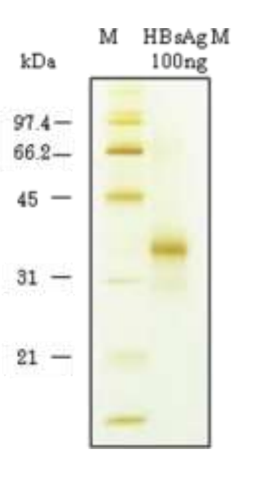
HBsAg, M-protein was subjected for SDS-PAGE (Silver staining) at 100ng.
Recently Viewed

No More Sales-NpFlamma® HGC ICG
$99999.00
Flamma® 648 Amine
$1100.80
Flamma® 488 Vinylsulfone
$3856.00
Flamma® 749 Amine
$1100.80


Reverse Transcription
$0.00
FSD Fluor™ 680 Phalloidin
$845.54
exsig™ COVID-19 Direct
$1873.00
Streptavidin, Flamma® 749
$443.68