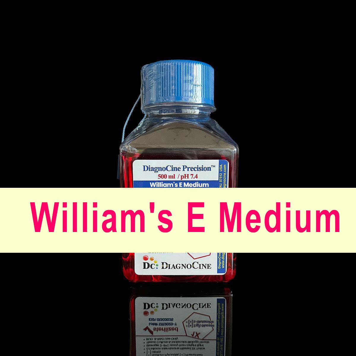
FluxMPS™ William's E Medium
Select WilliamsEMedium
- see the table below
FluxMPS™ William's E Medium
FluxMPS™ William's E Medium is a 1X liquid, ready-to-use formulation of the specialized cell culture medium originally developed by Williams and Gunn for long-term cultivation of adult rat liver epithelial cells. It is an enriched formulation that contains higher levels of amino acids and glucose compared to earlier media, and is notable for its unique ingredients, including zinc, iron, manganese, non-essential amino acids, glutathione as a reducing agent, and methyl linoleate lipid.
- 2 configurations: William's E (standard, with Sodium Bicarbonate), and William's E w/o Sodium Bicarbonate, each independently toggling L-Glutamine, Sodium Bicarbonate, HEPES, and Phenol Red
- This medium has proven versatile and effective for growing primary hepatocyte cells from various species, including human-derived HepaRG cells
- Typically requires supplementation with 5-10% fetal bovine serum (FBS), as it does not contain proteins or growth factors
- Uses a sodium bicarbonate buffer system (2.2 g/L) and needs a 5-10% CO2 environment to maintain physiological pH
- 2.0 g/L standard glucose concentration
- Its effectiveness in supporting long-term cell cultures and its applicability to various cell types have made it a valuable tool in cell biology research, particularly in liver cell studies
- Purified through FluxMPS™ quadruple-stage 0.1 micron / 0.04 micron filtration, engineered for organ-on-chip (OoC), tissue-on-chip (ToC), and lab-on-chip (LoC) microfluidic platforms
- Available in 500 mL and 1000 mL sizes; store at 2-8°C protected from light; every standard concentration and additive is customizable on request
- Concentration1X
- Glucose2.0 g/L
- BufferingSodium bicarbonate (2.2 g/L), 5-10% CO2
- Configurable supplementsL-Glutamine / Sodium Bicarbonate / HEPES / Phenol Red
- Unique ingredientsZinc, iron, manganese, glutathione, methyl linoleate
- FiltrationFluxMPS™ quadruple-stage, 0.04 micron final
- Sizes500 mL, 1000 mL
- Storage2-8°C, protected from light
At-a-Glance Supplement Matrix
Please select the supplement(s) of interest, then click Search. Every matching configuration lights up below. Click the catalog number or the View button to go straight to that product page.
| Name | Cat No. | L-Glutamine | Sodium Bicarbonate | HEPES | Phenol Red | Product Page |
|---|---|---|---|---|---|---|
| William's E Medium | DCP-WME1X | check | check | remove | check | Viewarrow_forward |
| William's E Medium, w/o Sodium Bicarbonate | DCP-WME-B1X | check | remove | remove | check | Viewarrow_forward |
William's Medium E: Defined Medium for Primary Hepatocyte Isolation and Long-Term Liver Cell Culture
William's Medium E is a chemically defined, synthetic basal medium developed between 1971 and 1974 by George M. Williams and colleagues, culminating in the 1974 publication by Williams and Gunn, which presented the formulation optimized for long-term primary culture of adult rat liver epithelial cells. It is the universally accepted default medium for primary hepatocyte culture, hepatic cell line maintenance, drug-metabolism studies, hepatotoxicity testing, and hepatocyte-based tissue engineering — roles that no Eagle-lineage medium (BME, MEM, DMEM, RPMI 1640) fulfills adequately without substantial supplementation. Its distinctiveness rests on a profile engineered around the metabolic, antioxidant, lipid, and micronutrient requirements of differentiated hepatic parenchymal cells: all four fat-soluble vitamins (A, D2, E, K3), a polyunsaturated fatty acid (methyl linoleate), four ultratrace transition-metal micronutrients (Fe, Cu, Mn, Zn), ascorbic acid, reduced glutathione, sodium pyruvate, a NEAA-rich 21-amino-acid profile, and a glucose concentration of 2000 mg/L (11.1 mM) more physiologically relevant to the liver than either low-glucose (5.5 mM) or high-glucose DMEM (25 mM).
Origins and Development
George M. Williams and the Liver Cell Culture Problem
Long-term primary culture of adult mammalian liver cells was a uniquely difficult challenge through the 1960s. Hepatic parenchymal cells (hepatocytes) are highly differentiated, non-dividing or slowly cycling cells with exceptionally demanding nutritional requirements arising from their role as the body's central metabolic organ. Unlike fibroblasts or lymphoid cells, which are sustained in Eagle-lineage or RPMI media, hepatocytes rapidly lost their differentiated functions (albumin secretion, cytochrome P450 activity, gluconeogenesis, urea synthesis) in all available media. Maintaining the differentiated phenotype required physiologically relevant glucose for gluconeogenic balance, a lipid source, all four fat-soluble vitamins, trace metals for liver-specific metalloenzymes, and antioxidant support adequate for a high-oxidative-metabolism cell type. George M. Williams, working at the Naylor Dana Institute for Disease Prevention (American Health Foundation, Valhalla, New York), addressed this through iterative modifications to the medium, beginning in 1971.
The 1971 Paper: Medium D and Epithelial Liver Cell Isolation
The developmental sequence begins with Williams, Weisburger & Weisburger (1971), "Isolation and long-term cell culture of epithelial-like cells from rat liver," Experimental Cell Research 69(1): 106-112 (PMID 5124481; DOI 10.1016/0014-4827(71)90316-8). Using a differential attachment / sequential plating technique, the group isolated epithelial-like cells from the livers of 10-day-old rats using a modified MEM called William's Medium D, enriched in amino acids and formulated with twice the glucose of standard MEM. Medium D was a transitional formulation demonstrating that enriched, defined media could better sustain liver epithelial cells than Eagle-lineage standards.
The 1974 Paper: William's Medium E
Williams and Gunn, working with adult rather than young rat liver cells and refining the nutritional provisions, released the definitive formulation: Williams & Gunn (1974), "Long-term cell culture of adult rat liver epithelial cells," Experimental Cell Research 89(1): 139-142 (PMID 4373256; DOI 10.1016/0014-4827(74)90196-7). This established William's Medium E as the standard for maintaining adult liver epithelial cells, demonstrating retention of epithelial morphology in primary culture, a prerequisite for any meaningful study of hepatocyte function.
Adoption for Primary Hepatocyte Culture
Following Per Ottar Seglen's two-step collagenase portal-vein perfusion method for high-yield hepatocyte isolation (1976), William's Medium E became the standard collagenase-perfusion vehicle and the default post-isolation plating and maintenance medium for primary rat and human hepatocytes. The combination of Seglen's isolation technique and William's Medium E defines the foundation of primary hepatocyte methodology still in use today, now routinely applied to hepatocytes from rat, mouse, human, pig, sheep, and other species.
William's Medium in the Classical Medium Family Tree
- Standard MEM (Eagle, 1959) — the Eagle-lineage baseline from which William's Medium D was modified.
- William's Medium D (Williams, Weisburger & Weisburger, 1971) — a modified MEM enriched in amino acids and formulated with twice the glucose of standard MEM, used to isolate epithelial-like cells from the livers of 10-day-old rats; a transitional formulation demonstrating that enriched, defined media could better sustain liver epithelial cells than Eagle-lineage standards.
- William's Medium E (Williams & Gunn, 1974) — the definitive formulation, refined for adult rat liver epithelial cells, establishing retention of epithelial morphology in primary culture and becoming the standard for maintaining adult liver epithelial cells.
- Adoption with Seglen's collagenase perfusion method (1976) — the combination of Seglen's two-step collagenase portal-vein perfusion isolation technique and William's Medium E defines the foundation of primary hepatocyte methodology still in use today, across rat, mouse, human, pig, sheep, and other species.
William's Medium E Standard Formulation Reference
The composition below reflects the canonical William's Medium E formulation (as in the Sigma and Gibco reference formulations and the original Williams & Gunn medium), cross-checked against the HiMedia AL125 datasheet. L-glutamine is supplied separately and added to a final concentration of 2 mM (292 mg/L) immediately before use. Quality control (HiMedia AL125 liquid): orangish-red clear solution; pH 7.00-7.60; osmolality 290-330 mOsm/kg; endotoxin NMT 1 EU/mL; 18-month shelf life. All values are as stated in the source; no additional numeric values are implied for any specific catalog number beyond what is stated here and in the supplement matrix above.
| Ingredient | mg/L |
|---|---|
| Calcium chloride dihydrate (CaCl2.2H2O) | 265.000 |
| Copper sulfate pentahydrate (CuSO4.5H2O) | 0.0001 |
| Ferric nitrate nonahydrate [Fe(NO3)3.9H2O] | 0.0001 |
| Magnesium sulfate, anhydrous (MgSO4) | 97.670 |
| Manganese chloride tetrahydrate (MnCl2.4H2O) | 0.0001 |
| Potassium chloride (KCl) | 400.000 |
| Sodium bicarbonate (NaHCO3) | 2200.000 |
| Sodium chloride (NaCl) | 6800.000 |
| Sodium phosphate monobasic, anhydrous (NaH2PO4) | 122.000 |
| Zinc sulfate heptahydrate (ZnSO4.7H2O) | 0.0002 |
| Amino Acid | mg/L |
|---|---|
| Glycine | 50.000 |
| L-Alanine | 90.000 |
| L-Arginine (free base) | 50.000 |
| L-Asparagine monohydrate | 20.000 |
| L-Aspartic acid | 30.000 |
| L-Cysteine | 40.000 |
| L-Cystine | 20.000 |
| L-Glutamic acid | 44.500 |
| L-Glutamine (add separately, 2 mM) | 292.000 |
| L-Histidine (free base) | 15.000 |
| L-Isoleucine | 50.000 |
| L-Leucine | 75.000 |
| L-Lysine hydrochloride | 87.460 |
| L-Methionine | 15.000 |
| L-Phenylalanine | 25.000 |
| L-Proline | 30.000 |
| L-Serine | 10.000 |
| L-Threonine | 40.000 |
| L-Tryptophan | 10.000 |
| L-Tyrosine disodium salt dihydrate | 50.000 |
| L-Valine | 50.000 |
| Vitamin | mg/L |
|---|---|
| Ascorbic acid (as sodium salt) | 2.000 |
| D-Biotin | 0.500 |
| Choline chloride | 1.500 |
| D-Calcium pantothenate | 1.000 |
| Ergocalciferol (Vitamin D2) | 0.100 |
| Folic acid | 1.000 |
| Menadione sodium bisulfite (Vitamin K3) | 0.010 |
| myo-Inositol | 2.000 |
| Niacinamide (Nicotinamide) | 1.000 |
| Pyridoxal hydrochloride (Vitamin B6) | 1.000 |
| Retinol acetate (Vitamin A) | 0.100 |
| Riboflavin (Vitamin B2) | 0.100 |
| Thiamine hydrochloride (Vitamin B1) | 1.000 |
| alpha-Tocopherol phosphate sodium salt (Vitamin E) | 0.010 |
| Vitamin B12 (Cyanocobalamin) | 0.200 |
| Other Components | Amount |
|---|---|
| D-Glucose (Dextrose) | 2000.000 mg/L |
| Glutathione (reduced, GSH) | 0.050 mg/L |
| Methyl linoleate | 0.030 mg/L |
| Phenol red (sodium salt) | 10.000 mg/L |
| Sodium pyruvate | 25.000 mg/L |
William's Medium E uses the MEM Earle's salt base (NaCl, CaCl2, MgSO4, KCl, NaH2PO4, NaHCO3) but extends it with four ultratrace transition-metal micronutrients: Fe, Cu, Mn, and Zn. This trace-element set is the defining inorganic signature of William's Medium E and is absent from the Eagle-lineage media and RPMI 1640. Copper, manganese, and zinc are absent from BME, MEM, DMEM, and RPMI 1640 — the unique William's E trio. L-Glutamine is not included in the standard liquid formulation and must be added freshly to 2 mM (292 mg/L) before use; the exclusion is intentional, since glutamine degrades during storage to pyroglutamate and ammonia, so the medium is supplied glutamine-free to preserve shelf life.
William's Medium E vs. Classical Media
| Feature | William's Medium E | DMEM (High-Glucose) | RPMI 1640 | Medium 199 | Waymouth MB 752/1 |
|---|---|---|---|---|---|
| Developer & year | G.M. Williams & Gunn - 1974 | Dulbecco & Freeman, 1959 | Moore et al., 1966/1967 | Morgan, Morton & Parker, 1950 | Charity Waymouth, 1959 |
| Primary design goal | Primary hepatocyte long-term culture | Adherent monolayer culture | Suspension lymphoid culture | Universal comprehensive defined | Serum-free L929 fibroblast |
| Amino acids (entries) | 21 (dual cysteine + cystine) | 15 | 20 | 21 | 18 |
| Vitamins (count) | 15 (all four fat-soluble) | 8 | 11 | 17 | 11 |
| Vitamin A (retinol acetate) | 0.100 mg/L | Absent | Absent | 0.140 mg/L | Absent |
| Vitamin D2 (ergocalciferol) | 0.100 mg/L | Absent | Absent | 0.100 mg/L | Absent |
| Vitamin E (alpha-tocopherol phosphate) | 0.010 mg/L | Absent | Absent | 0.010 mg/L | Absent |
| Vitamin K3 (menadione) | 0.010 mg/L | Absent | Absent | 0.016 mg/L | Absent |
| Ascorbic acid | 2.000 mg/L | Absent | Absent | 0.050 mg/L | 17.500 mg/L |
| Vitamin B12 | 0.200 mg/L | Absent | 0.005 mg/L | Absent | 0.200 mg/L |
| Biotin | 0.500 mg/L | Absent | 0.200 mg/L | 0.010 mg/L | 0.020 mg/L |
| Pyridoxal HCl (B6 form) | Pyridoxal HCl | Pyridoxine HCl | Pyridoxine HCl | Both forms | Pyridoxine HCl |
| Trace metals (Cu, Mn, Zn) | Present | Absent | Absent | Absent (Fe only) | Absent |
| Methyl linoleate (lipid) | 0.030 mg/L | Absent | Absent | Cholesterol 0.2 + Tween 80 | Absent |
| Reduced glutathione | 0.050 mg/L | Absent | 1.000 mg/L | 0.050 mg/L | 15.000 mg/L |
| Sodium pyruvate | 25.000 mg/L | Optional (110 mg/L) | Absent (standard) | Absent | Absent |
| Glucose | 2000 mg/L (~11.1 mM) | 4500 mg/L (25 mM) | 2000 mg/L (11.1 mM) | 1000 mg/L (5.5 mM) | 5000 mg/L (27.8 mM) |
| NaHCO3 | 2200 mg/L | 3700 mg/L | 2000 mg/L | 2200 mg/L | 2240 mg/L |
| pH (with NaHCO3) | 7.00-7.60 | ~7.2 | 7.0-7.4 | 7.2 | 7.1-7.7 |
| Osmolality (with NaHCO3) | 290-330 mOsm/kg | 320-355 mOsm/kg | 255-310 mOsm/kg | 310-350 mOsm/kg | 305-345 mOsm/kg |
| Calcium source | CaCl2.2H2O 265 mg/L | CaCl2.2H2O 265 mg/L | Ca(NO3)2.4H2O 100 mg/L | CaCl2.2H2O 265 mg/L | CaCl2.2H2O 120 mg/L |
| Iron source | Fe(NO3)3.9H2O (ultratrace) | Fe(NO3)3.9H2O 0.1 mg/L | None | Fe(NO3)3.9H2O 0.72 mg/L | None |
| Preformed purines | None | None | None | Adenine, hypoxanthine | Hypoxanthine |
| Fat-soluble vitamins | A, D2, E, K3 (all four) | None | None | A, D2, E, K3 (all four) | None |
| L-Glutamine supplied | Separately (add 2 mM fresh) | In standard formulation | In standard formulation | In standard formulation | In standard formulation |
| Primary validated cells | Primary hepatocytes, HepaRG | HEK293, MEFs | Jurkat, PBMCs, hybridomas | Chick fibroblasts, oocytes | L929 (serum-free) |
William's Medium E: G.M. Williams & Gunn, 1974. DMEM: Dulbecco & Freeman 1959. RPMI 1640: Moore et al., 1966/1967. Medium 199: Morgan, Morton & Parker, 1950. Waymouth MB 752/1: Charity Waymouth, 1959.
Why FluxMPS™
Quadruple-Stage Purity
Every FluxMPS™ William's E configuration is purified to 0.04 microns — finer than any ready-to-use cell culture media currently available — before it ever reaches your chip.
Engineered for Microfluidics
Designed from the ground up for Organ-on-Chip (OoC), Tissue-on-Chip (ToC), and Lab-on-Chip (LoC) platforms, where the medium itself is part of the instrument.
Particulate & Aggregate Removal
The quadruple-stage architecture eliminates microscopic particulates and protein aggregates that silently block micro-channels and disrupt laminar flow — a particular concern for collagen-coated, hepatocyte-specific microfluidic cultures.
Optical Clarity
Optical clarity supports real-time imaging and integrated biosensing on Organ-on-Chip platforms without background interference.
Regulatory-Aligned Foundation
Formulated to support FDA-recognized physiological modeling standards, providing a validated, reproducible media foundation as downstream data moves toward regulatory scrutiny.
Translational Research Ready
A single, consistent media platform supporting drug discovery, toxicology screening, and translational research from bench through preclinical modeling.
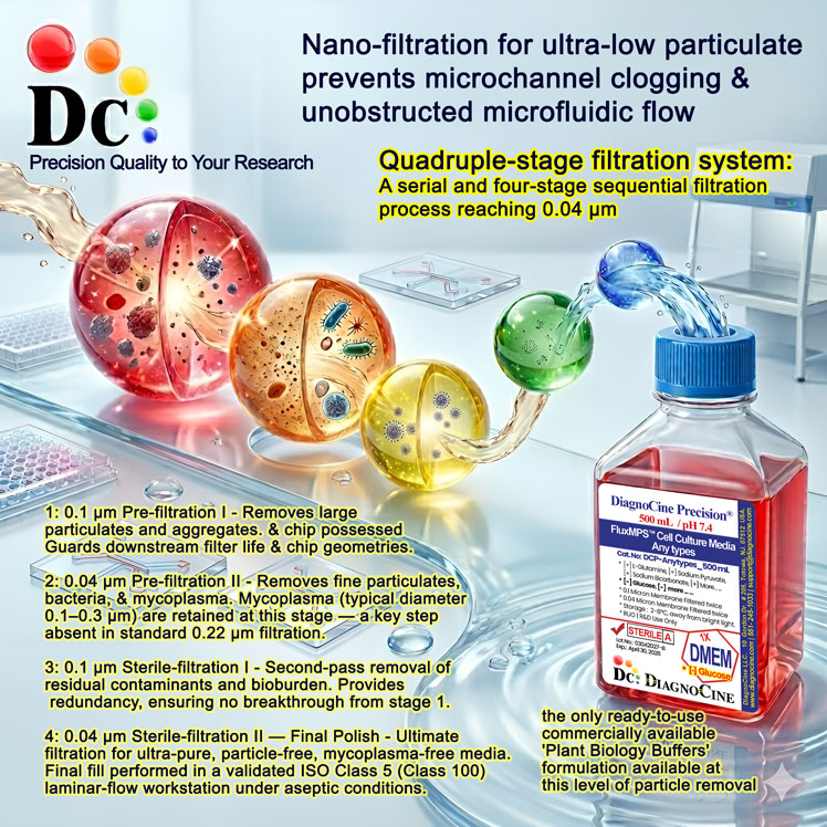
Quadruple-Stage Filtration
Every FluxMPS™ William's E batch passes through a four-stage filtration train before final fill, engineered specifically for microfluidic cell culture applications.
-
1
0.1 µmPre-filtration Stage One
Bulk particulate reduction prior to sterile filtration.
-
2
0.1 µmPre-filtration Stage Two
Second-pass pre-filtration to protect the downstream sterile filters and extend their working life.
-
3
0.04 µmSterile Filtration Stage One
First sterile-filtration pass at 0.04 microns — finer than any ready-to-use cell culture media currently available.
-
4
0.04 µmSterile Filtration Stage Two
Second sterile-filtration pass, eliminating microscopic particulates and protein aggregates before final fill.
Built for Continuous Flow
Zero-clogging performance across complex micro-channel geometries and long-term automated perfusion studies running continuously for weeks.
Validated Cell Lines & Applications
This medium has proven versatile and effective for growing primary hepatocyte cells from various species, including human-derived HepaRG cells. Its effectiveness in supporting long-term cell cultures and its applicability to various cell types have made it a valuable tool in cell biology research, particularly in liver cell studies.
Primary Hepatocyte Isolation & Culture
William's Medium E is the globally established standard medium for every stage of primary hepatocyte work: collagenase perfusion isolation, post-digestion washing and Percoll gradient separation, and post-isolation culture on collagen-I-coated plates with serum, insulin, and dexamethasone or hydrocortisone. Species validated include rat, mouse (C57BL/6), human (resection specimens), pig, sheep, and zebrafish.
HepaRG Cells
HepaRG is a bipotent human hepatoma progenitor line that differentiates into hepatocyte-like cells expressing CYP3A4, CYP2E1, and albumin. William's Medium E is the standard base medium for all stages of HepaRG culture, via a proliferation phase followed by a 2% DMSO differentiation phase, producing the highest expression of liver-specific enzymes and characteristic bile-canaliculi morphology.
HepG2 & Huh7
HepG2 (hepatocellular carcinoma) and Huh7 (hepatoma) are sometimes cultured in William's Medium E for experiments requiring a liver-appropriate medium, though DMEM + 10% FBS is their more common routine maintenance medium. For hepatocyte-specific drug- or lipid-metabolism experiments, William's Medium E avoids the confounding effects of DMEM's supraphysiologic glucose on lipogenic enzyme induction.
iPSC Hepatocyte Differentiation
A 2016 study demonstrated improved survival and initiation of hepatocyte differentiation when human iPSCs were shifted from pluripotency media to William's Medium E prior to hepatocyte-differentiation-inducer treatment, establishing the medium as a transitional bridge between pluripotency maintenance and hepatocyte identity induction.
Drug Metabolism & Cytochrome P450 Studies
William's Medium E is the standard medium for in vitro drug metabolism studies with primary hepatocytes in regulatory (OECD, ICH) and pre-regulatory settings. CYP-induction testing maintains primary hepatocytes in William's Medium E for 24-72 hours before inducer exposure; the EURL-ECVAM DB-ALM protocol for CYP induction specifies William's Medium E as the culture medium for validated primary hepatocyte CYP induction assays.
Chemical Carcinogenesis, Hepatotoxicity & Progenitor Lines
Rat liver epithelial cells in William's Medium E respond appropriately to procarcinogens requiring enzymatic activation, and primary rat and human hepatocytes in William's Medium E are used for acute and repeated-dose hepatotoxicity assessment. Liver progenitor lines (e.g., BC2/Bac2) are maintained in William's Medium E + serum + insulin + hydrocortisone hemisuccinate, and differentiated HepaRG cells in William's Medium E + DMSO serve as a primary in vitro hepatitis B virus entry/replication model.
Practical Considerations
L-Glutamine Instability
William's Medium E is supplied without L-glutamine because glutamine hydrolyzes to pyroglutamate and ammonia during storage. For primary hepatocytes, this matters acutely: hepatocytes are the primary site of ammonia detoxification, and ammonia levels above ~1 mM inhibit urea-cycle enzyme expression. Add L-glutamine freshly at 2 mM and use within approximately 4 weeks of addition.
DMSO in HepaRG Differentiation & Collagen Coating
The 2% DMSO added during HepaRG differentiation is a functional component, not a cryoprotectant: at this concentration, it activates CAR and PXR, driving hepatocyte-specific gene programs including CYP induction. Unlike DMEM-supported lines, primary hepatocytes and HepaRG cells in William's Medium E require collagen-I or Matrigel coating for attachment; uncoated surfaces cause rounding, loss of polarity, and accelerated de-differentiation even in fully supplemented medium.
Serum-Free Use
William's Medium E can be used serum-free for primary hepatocytes when supplemented with insulin, transferrin, selenium, dexamethasone or hydrocortisone, and free fatty acids bound to delipidated albumin; chemically defined, serum-free William's E formulations are commercially available for GMP-compliant applications.
Light Sensitivity
Retinol acetate, riboflavin, and methyl linoleate make William's Medium E more photosensitive than DMEM or RPMI 1640; follow amber-vessel/foil storage and minimize light exposure.
Frequently Asked Questions
Verified References
- Williams, G.M., Weisburger, E.K. & Weisburger, J.H. (1971). Isolation and long-term cell culture of epithelial-like cells from rat liver. Experimental Cell Research, 69(1): 106-112. PMID 5124481. DOI 10.1016/0014-4827(71)90316-8.
- Williams, G.M. & Gunn, J.M. (1974). Long-term cell culture of adult rat liver epithelial cells. Experimental Cell Research, 89(1): 139-142. PMID 4373256. DOI 10.1016/0014-4827(74)90196-7.
- Williams, G.M. (1976). Primary and long-term culture of adult rat liver epithelial cells. Methods in Cell Biology, 14: 357-364.
- Laishes, B.A. & Williams, G.M. (1976). Conditions affecting primary cell cultures of functional adult rat hepatocytes. I. The effect of insulin. In Vitro, 12: 521-532.
- Seglen, P.O. (1976). Preparation of isolated rat liver cells. Methods in Cell Biology, 13: 29-83.
- Guillouzo, A., et al. (2007). The human hepatoma HepaRG cells: a highly differentiated model for studies of liver metabolism and toxicity of xenobiotics. Chemico-Biological Interactions, 168(1): 66-73.
- Yao, T. & Asayama, Y. (2017). Animal-cell culture media: history, characteristics, and current issues. Reproductive Medicine and Biology, 16(2): 99-117. PMID 29259457. PMC5661806.
FluxMPS™ — Precision Cell Culture Media for Microphysiological Systems
Built for the architecture of the future. Not the flask of the past.
Traditional cell culture media were formulated for static well plates and flasks — environments that tolerate impurities, precipitates, and particle loads that would immediately compromise a microfluidic system. FluxMPS™ was designed from the ground up for Organ-on-Chip (OoC), Tissue-on-Chip (ToC), and Lab-on-Chip (LoC) platforms, where the medium itself is part of the instrument.
Purity That Protects Your Platform
FluxMPS™ is purified to 0.04 microns — finer than any ready-to-use cell culture media currently available. At this level, the microscopic particulates and protein aggregates that silently block micro-channels, disrupt laminar flow, and generate false biological signals are eliminated before the media ever reaches your chip.
The result: your platform stays operational, your data stays clean, and your biology drives the result — not your media.
Engineered for Flow, Not Just Growth
The name FluxMPS™ reflects its core design principle. Every component is optimized for consistent, laminar flow performance across:
- Complex micro-channel geometries
- Capillary-bed and vascular simulations
- Long-term automated perfusion studies running continuously for weeks
Zero-clogging performance is not a feature — it is the baseline specification.
Applications & Performance
| Application | What FluxMPS™ Delivers |
|---|---|
| Microfluidics | Stable shear stress; no channel blockage |
| Metabolic Tracing | Ultra-pure matrix with no contaminant interference |
| Long-term Perfusion | Consistent formulation stability over weeks of continuous flow |
| Organ-on-Chip | Optical clarity for real-time imaging and integrated biosensing |
Regulatory Foundation
FluxMPS™ is formulated to support FDA-recognized physiological modeling standards, providing a validated, reproducible media foundation for drug discovery, toxicology screening, and translational research. When your downstream data needs to stand up to regulatory scrutiny, your upstream media cannot be an afterthought.
The Bottom Line
Microfluidic platforms are precision instruments. They require precision inputs.
FluxMPS™ is the only ready-to-use cell culture medium engineered specifically to meet that standard — protecting your chip, your cells, and your science.