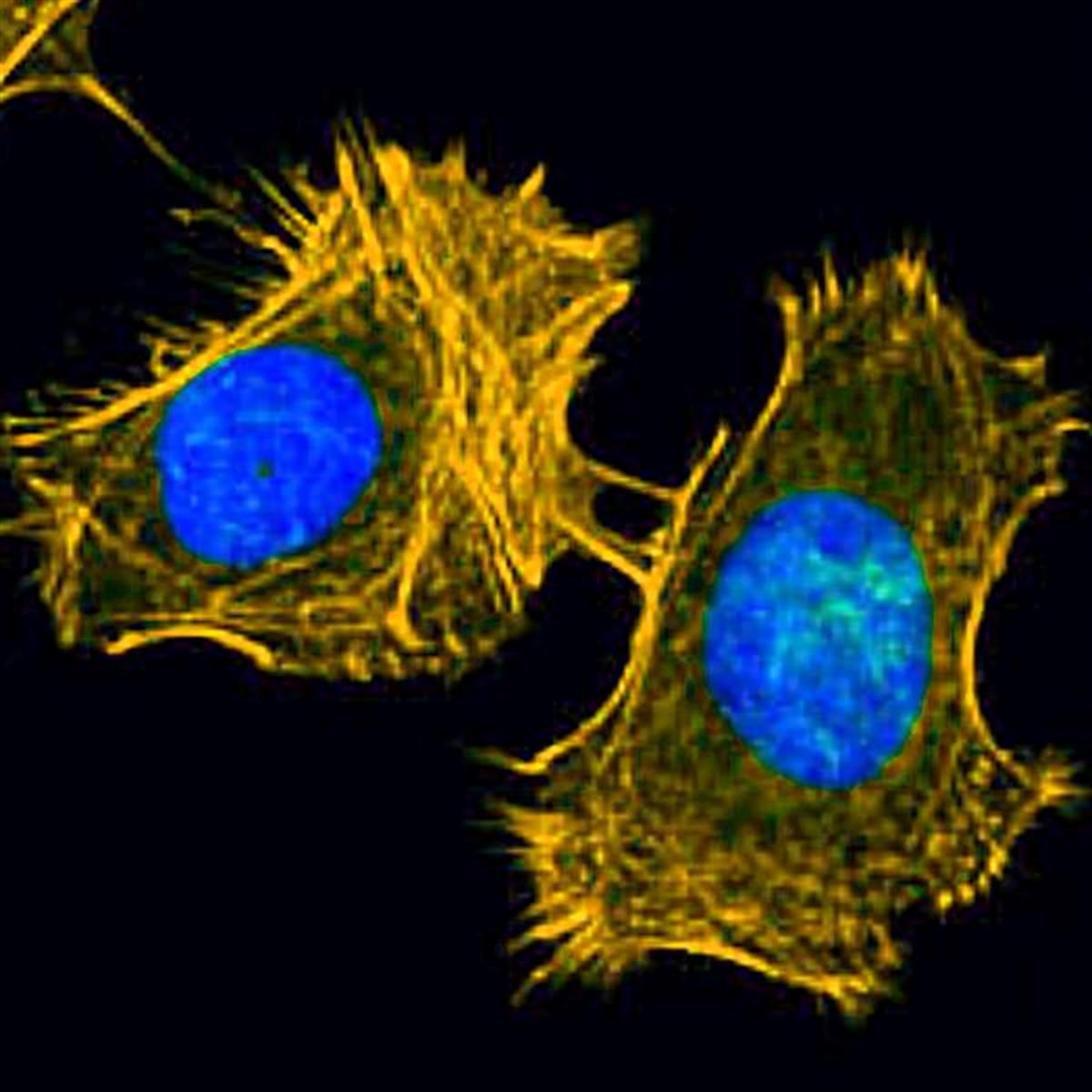
FSD Fluor™ 555 Phalloidin
FSD Fluor™ 555 Phalloidin
Cat. No. List below
Description
Key Features:
1. High specificity for F-actin
2. Bright and photostable fluorescence
3. Stoichiometric binding to actin filaments
4. Excellent signal-to-noise ratio
5. Compatible with fixed and permeabilized samples
Applications:
1. Visualization of F-actin in formaldehyde-fixed and permeabilized tissue sections
2. Cytoskeleton imaging in cell cultures
3. Quantitative analysis of F-actin in cell-free experiments
4. Study of actin dynamics and cellular morphology
5. Detection of cells with compromised membranes
6. Super-resolution microscopy techniques like SIM and STORM
7. Fluorescence correlation spectroscopy
8. Fluorescence polarization measurements
Advantages:
1. Superior brightness and photostability compared to traditional fluorophores
2. Low background fluorescence due to selective F-actin binding
3. Versatile use across various cell types and species
4. Enables detailed imaging of actin structures
5. Ideal for both widefield and confocal microscopy techniques
While FSD Fluor™ 555 Phalloidin is a powerful tool for F-actin visualization, it is important to note that it is not cell-permeable and should be used with fixed and permeabilized samples. However, it may penetrate the membranes of certain hypoxic cells. For optimal results, use in formaldehyde-fixed and permeabilized tissue sections, cell cultures, or cell-free systems.
Specifications
- Fluorophore: FSD Fluor™ 555
- Application: Actin, Cytoskeleton imaging
- Excitation/Emission Max.(nm): 554/565 nm
- Storage conditions: -20 ℃, protect from light
Table 1. List of FSD Fluor™ Phalloidin
| Quick link (Cat.#) | Series | λEx (nm) | λEm (nm) | Packing unit |
| RCS2114 | FSD Fluor™ 488 Phalloidin | 495 | 519 | 300 tests |
| RCS2214 | FSD Fluor™ 555 Phalloidin | 554 | 565 | 300 tests |
| RCS2314 | FSD Fluor™ 594 Phalloidin | 593 | 618 | 300 tests |
| RCS2414 | FSD Fluor™ 647 Phalloidin | 651 | 667 | 300 tests |
| RCS2514 | FSD Fluor™ 680 Phalloidin | 679 | 696 | 300 tests |
| RCS2614 | FSD Fluor™ 750 Phalloidin | 751 | 774 | 300 tests |
| RCS2714 | FSD Fluor™ 800 Phalloidin | 774 | 790 | 300 tests |
Phalloidin is a rigid bicyclic peptide toxin isolated from Amanita phalloides mushroom that commonly used in imaging applications to selective label F-actin. Phalloidin is known to react stoichiometrically with actin, strongly promote actin polymerization, and stabilize actin polymers. Due to the ability to bind F-actin selectively, fluorescence conjugated phalloidin derivatives are widely used in microscopy for investigating the distribution of F-actin in cells. In biomedical research, fluorescent phalloidin probes are utilized in localizing actin filaments in living or fixed cells as well as for visualizing individual actin filaments in vitro. Fluorescent phalloidins much smaller than fluorescent antibodies, thus they have several advantages for actin labeling such as virtually identical binding properties with actin from different species of plants and animals, much denser labeling of filamentous actin, more detailed images, and lower nonspecific binding. Fluorescent phalloidin conjugates are not permeable to most live cells, thus they should be used to detect cells with compromised membranes. However, fluorescent labeled phalloidins may penetrate the membranes of certain hypoxic cells. BioActs offers FSD Fluor™ Phalloidin series for the labeling and quantitative analysis of F-actin in formaldehyde-fixed and permeabilized tissue sections, cell cultures, and cell-free experiments.
Actin Labeling Protocol
Materials Required but Not Provided
- Methanol
- 1X Phosphate buffered saline (PBS)
- 4% Paraformaldehyde in PBS
- Triton X-100
- 0.1% PBST
- 2% BSA in 0.1% PBST
- Cell line prepared in a culture container suitable for imaging
- The appropriate cell culture medium for the cell line
- Micropipettes
- Fluorescence microscope
- Cell incubator (37 °C)
- Seed the cells in confocal dish by adequate amount to prepare stabilized cells.
- Wash the cells twice with pre-warmed PBS at 37°C
- Treat cells with 4% paraformaldehyde in PBS for 10 min to fix the cells.
- Wash the cells twice with PBS.
- Treat 0.1% Triton X-100 in PBS (0.1% PBST) for 10 min. at room temperature for permeabilization.
- Wash the cell twice with PBS.
- Add 2% BSA in 0.1% PBST for 30 minutes at room temperature to perform the blocking.
- Dilute 5uL of methanolic stock solution of FSD Fluor™ Phalloidin into 2% BSA in 0.1% PBST (1mL) and staining for 1 h at room temperature. - The amount of dye and staining time are inversely proportional
- Wash the cell twice with PBS.
Additional Mounting for long-term storage
- Remove PBS from the last step.
- Treat mounting solution onto of the cells.
- Seal the edge of the coverslip with nail polish and store the sample in the dark at 2–6°C.
Citation & Reference
1. Lynen F, Wieland U (18 November 1937). "Uber die Giftstoffe des Knollenblätterpilzes. IV". Justus Liebigs Annalen der Chemie (in German). 533 (1): 93–117.
2. Wieland T, Schon W (16 January 1955). "Über die Giftstoffe des grünen Knollenblätterpilzes X. Mitteilung. Die Konstitution des Phalloidins". Justus Liebigs Annalen der Chemie. 593 (2): 157–178.
3. Immunohistochemistry: Basics and Methods. Springer Science & Business Media. 2010. pp. 92–3.
4. Walton JD; Hallen-Adams He; Luo H (4 August 2010). "Ribosomal biosynthesis of the cyclic peptide toxins of Amanita mushrooms". Peptide Science. 94 (5): 659–654.
5. Anderson MO, Shelat AA, Kiplan Guy R (16 April 2005). "A solid-phase approach to the phallotoxins: total synthesis of [ala7]-phalloidin". J. Org. Chem. 70 (12): 4578–84.
6. Wieland T (1963). "Chemical and toxicological studies with cyclopeptides of Amanita phalloides". Pure and Applied Chemistry. 3 (6): 339–350.
7. Schröder, Eberhard; Lübke, Klaus (2014). The Peptides, Volume II: Synthesis, Occurrence, and Action of Biologically Active Polypeptides. Elsevier. p. 475.
8. Cooper JA (October 1987). "Effects of cytochalasin and phalloidin on actin". J. Cell Biol. 105 (4): 1473–8.
9. Barden JA, Miki M, Hambly BD, Dos Remedios CG (February 1987). "Localization of the phalloidin and nucleotide-binding sites on actin". Eur. J. Biochem. 162 (3): 583–8.
10. Wehland J, Osborn M, Weber K (December 1977). "Phalloidin-induced actin polymerization in the cytoplasm of cultured cells interferes with cell locomotion and growth". Proc. Natl. Acad. Sci. U.S.A. 74 (12): 5613–7.
11. Capani F, Deerinck TJ, Ellisman MH, Bushong E, Bobik M, Martone ME (1 November 2001). "Phalloidin-eosin followed by photo-oxidation: a novel method for localizing F-actin at the light and electron microscopic levels". J. Histochem.Cytochem. 49 (11): 1351–61.