

Anti-p53(pSer46), Phosphorylated, Mouse-Mono(36)
Product#: FNK-71-115
$346.79
Availability:
Ships in 24 hours
Anti-p53(pSer46), Phosphorylated, Mouse-Mono(36)
General information
| Cat. No. | :FNK-71-115 |
| Size | :100 ug |
| Host Species | :Mouse |
| Reactivity | :Human p53-phosphorylated at Ser46. Not tested in other species. |
| Immunogen | :Synthetic peptide containing phospho-Ser46 of p53 |
| Application |
|
| Isotype | :mouse IgG1 (κ) |
| Product | :Purified from serum-free culture medium of hybridoma (#36) by proprietary chromatography procedures under mild conditions. |
| Storage | :Sent at 4℃ or -20 ℃ and store at -20℃. Do not freeze |
| Form | :1mg/ml in PBS, 50% glycerol, filter-sterilized. Azide- and carrier-free. |
| Data Link | :UniProtKB/Swiss-Prot P04637 (P53_HUMAN) |
Description
p53 mutants are found in more than half of human cancers and are considered as the most important human cancer related gene. p53 is detected at 53kD position by electrophoresis and is composed of 393 amino acids. In the unstressed normal cells the p53 level is low and it is inactive. However, with stress, especially with DNA damage, it is activated to promote arrest of cell cycle and repair of DNA damage, or induction of apoptosis. The functions of p53 are regulated by phosphorylation of serine and threonine, and acetylation of lysine at various sites in the molecule. Among the phosphorylation sites, Ser46 is phosphorylated when DNA damage is so severe as to become unrepairable, which leads to apoptosis by activating transcription of proapoptotic genes such as p53AIPI As to the kinase of phosphorylation of Ser46, involvement of DYRK2 and ATM have been implicated
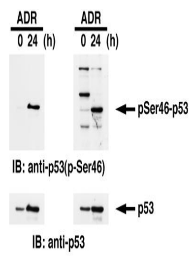 |
Fig.1. Identification of Ser46-phosphorylated p53 protein by western blotting. Samples: Crude cell extracts of MOLT-4 untreated (left lanes) and treated with adriamycin for 24 h (right lanes). The left panel is the result with our product and the right panel is the one obtained with the product of our competitor. The lower panel is the whole p53 protein identified by omnipotent anti-p53 antibody |
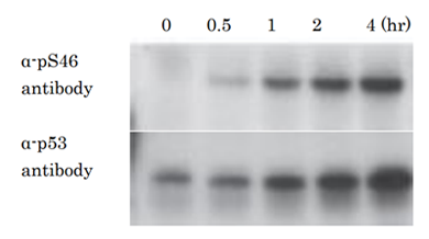 |
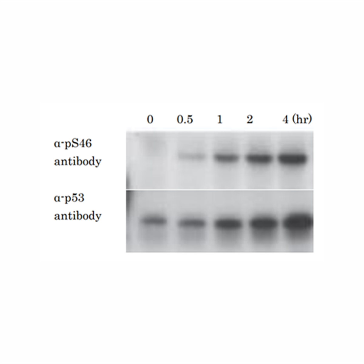
Fig.2. Kinetics of phosphorylation of p53 at Ser46 after X-ray irradiation .
Samples of U2OS cells (human osteosarcoma) were taken at the indicated times after X-ray irradiation at 10 Gy and analyzed by western blotting with anti-p53 p-S46 antibody (#36) and anti-p53 antibody (non BioAcademia). Primary antibodies were diluted with “ Can Get Signal “ signal enhancer (Toyobo, Osaka). |
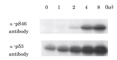 |
Fig.3. Kinetics of phosphorylation of p53 at Ser46 after UV-irradiation.
Samples of MCF7 cells (human beast cancer cell line) were taken at the indicated times after UV-irradiation at 20 J/m2 and analyzed by western blotting with anti-p53 p-S46 antibody (#36) and anti-p53 antibody (non BioAcademia).Primary antibodies were diluted with “ Can Get Signal “ signal enhancer |
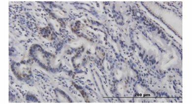 |
Fig.4. Immunohistochemistry of stomach cancer.
|
References
This product was used in the following publications.
- .Taira N. et al (2007) DYRK2 is targeted to the nucleus and controls p53 via Ser46 phosphorylation in the apoptotic response to DNA damage. Mol. Cell 25:725-738. PMID: 17349958 WB
- Kodama M. et al (2010) Requirement of ATM for rapid p53 phosphorylation at Ser46 without Ser/Thr-Gln sequences. Mol Cell Biol. 30:1620-33. PMID20123963. WB
- Taira N. et al (2010) ATM augments nuclear stabilization of DYRK2 by inhibiting MDM2 in the apoptotic response to DNA damage. J Biol Chem. 285:4909-19. PMID: 19965871 WB

Recently Viewed

Western Marker Neo (wide)
$348.95




Flamma® 552 PEG4-Alkyne
$1632.80
Goat anti-rabbit IgG, FSD™ 647
$339.20

Beauvericin, 1 mg
$87.72