Anti-SUMO-1 antibody, rat monoclonal (4D12), Biotin-conjugated
Product#: FNK-70-654
$191.84
Availability:
Ships in 1-2 Weeks
Anti-SUMO-1 antibody, rat monoclonal (4D12), Biotin-conjugated
Product ID and Size
| Cat. No. | Size | Labeled | Data Link | Link |
| FNK-70-653 | 100 µg | Unlabeled | Swiss-Prot P63165 |
 |
| FNK-70-654 | 50 µg | Biotinylated | Swiss-Prot P63165 |
|
| FNK-70-655 | 50 µg | FITC-conjugated | Swiss-Prot P63165 |
|
General information
| Cat. No. | :70-654 |
| Size | :50 µg |
| Label | :Biotin |
| Antigen Species | :Human |
| Host Species | :Rat |
| Specificity | :Specific to human, simian, mouse and rat SUMO1. Other species have not been tested. |
| Immunogen | :Recombinant GST-fused human SUMO-1 (full length) |
| Product | :The antibody was produced in serum-free medium and purified by proprietary chromatography procedures under mild conditions and conjugated with biotin. |
| Isotype | :Rat IgG 2a kappa |
| Form | :1mg/ml in PBS, 50% glycerol, filter-sterilized. Azide- and carrier protein-free. |
| Application | 1. Western blotting (1/1,000 dilution) 2. Immunofluorescence staining (1/100 dilution) 3. Immunohistochemistry, frozen section (1/100 dilution) 4. ELISA (assay dependent) Other applications have not been tested. |
| Cross Reactivity | :Human/Mouse/Rat |
| Storage | :Shipped at 4℃ or -20℃ and store at -20℃. Do not freeze. |
| Data Link | :Swiss-Prot P63165 (human) |
Description
SUMO (Small Ubiquitin-like Modifier) proteins are a family of small proteins that are covalently attached to and detached from other proteins in cells to modify their function. Unlike ubiquitination which targets proteins for degradation SUMO modification plays a critical role in a number of cellular functions including nucleocytoplasmic transport gene expression cell cycle and formation of subnuclear structures such as promyelocytic leukemia (PML) bodies.
There are three confirmed SUMO isoforms in human; SUMO1 SUMO2 and SUMO3. SUMO2 /3 show a high degree of similarity to each other and are distinct from SUMO-1. Individual SUMO family members are all targeted to different proteins with diverse biological functions. SUMO-1 is conjugated to RanGAP PML p53 and IκB-α to regulate nuclear trafficking formation of subnuclear structures regulation of transcriptional activity and protein stability. SUMO1 is encoded as a 101 aa protein and first Met and C-terminal 4 aa are removed from the preprotein.
There are three confirmed SUMO isoforms in human; SUMO1 SUMO2 and SUMO3. SUMO2 /3 show a high degree of similarity to each other and are distinct from SUMO-1. Individual SUMO family members are all targeted to different proteins with diverse biological functions. SUMO-1 is conjugated to RanGAP PML p53 and IκB-α to regulate nuclear trafficking formation of subnuclear structures regulation of transcriptional activity and protein stability. SUMO1 is encoded as a 101 aa protein and first Met and C-terminal 4 aa are removed from the preprotein.
References: This antibody was used in Ref. 3 and 4.
1. Ulrich HD "The fast-growing business of SUMO chains.” Review Mol Cell 32: 301–305 (2008) PMID: 18995828
2. Cheng J et al “Role of desumoylation in the development of prostate cancer.” Review Neoplasia 8: 667-676 (2006) PMID: 16925949
3. Uchimura Y et al “Involvement of SUMO modification in MBD1- and MCAF1-mediated heterochromatin formation.” J Biol Chem 281: 23180-23190 (2006) PMID: 16757475
4. Saitoh N et al “In situ SUMOylation analysis reveals a modulatory role of RanBP2 in the nuclear rim and PML bodies.” Exp Cell Res 312: 1418-1430 (2006) PMID: 16688858
2. Cheng J et al “Role of desumoylation in the development of prostate cancer.” Review Neoplasia 8: 667-676 (2006) PMID: 16925949
3. Uchimura Y et al “Involvement of SUMO modification in MBD1- and MCAF1-mediated heterochromatin formation.” J Biol Chem 281: 23180-23190 (2006) PMID: 16757475
4. Saitoh N et al “In situ SUMOylation analysis reveals a modulatory role of RanBP2 in the nuclear rim and PML bodies.” Exp Cell Res 312: 1418-1430 (2006) PMID: 16688858
Figures
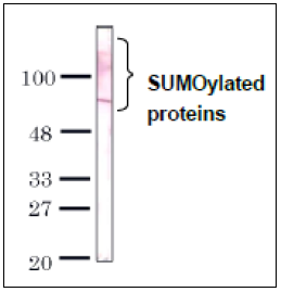 |
Fig.1 Detection of SUMO-1 by Western blotting with the antibody 4D12. An 80 kDa single and other multiple bands were observed in HeLa total cell extract. The 80 kDa band would be SUMO-RanGAP. Anti-SUMO-1 antibody 4D12 was used at 1 μg/ml. |
|
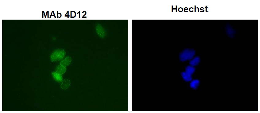 |
Fig.2 Immunofluorescence staining of SUMO-1 with the antibody 4D12 in the mouse primary culture neurons. Left: Stained with anti-SUMO-1 antibody 4D12 at 10 μg/ml. Right: DNA was stained with Hoechst |
|
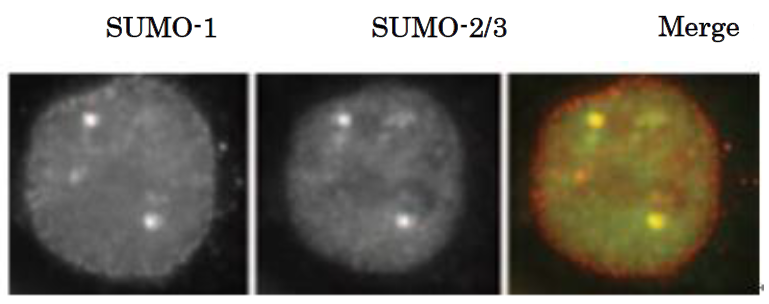 |
Fig.3 SUMO-1 colocalizes with SUMO2/3 as revealed by indirect immunofluorescence staing of C-33A cells (human cervix carcinoma). Left: SUMO-1 was stained withanti-SUMO1 antibody (4D12) at 10 μg/ml. Middle: SUMO2/3 was stained with anti-SUMO2/3 antibody (3H12) at 10 μg/ml. Right: Merged image |
|
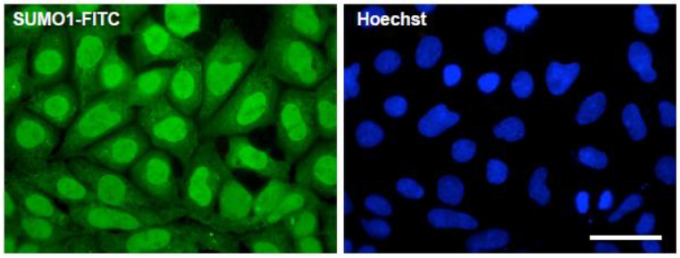 |
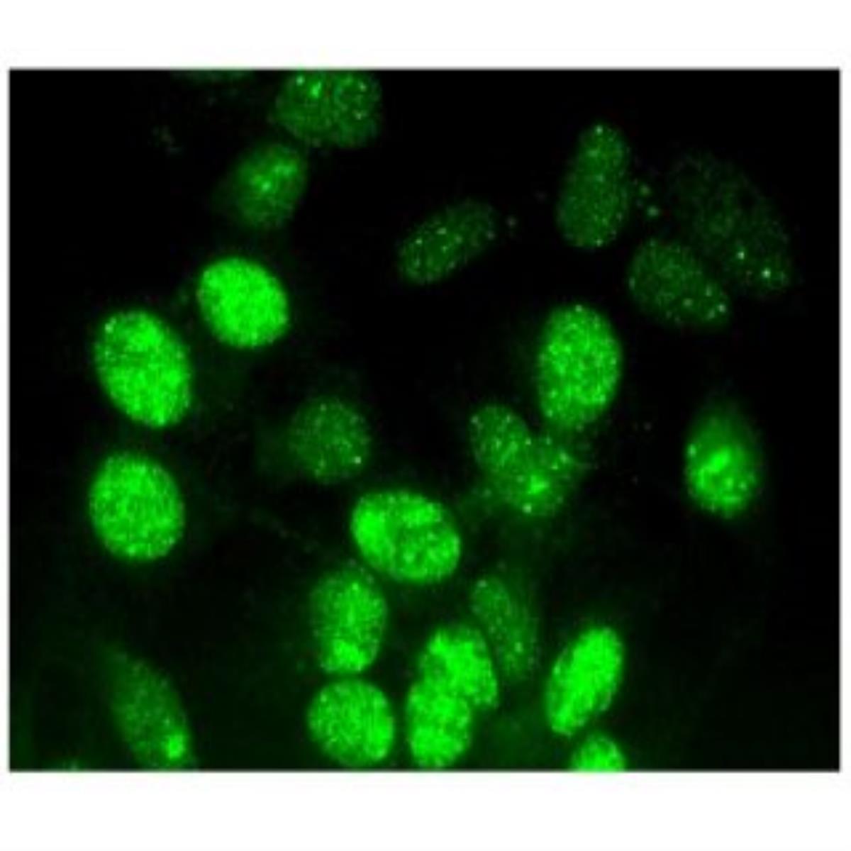
Fig.4 Fluorescence immunocytochemistry for endogenous SUMO1 expression. HEK293A cells were fixed, permeabilized, and stained with SUMO1-FITC (1: 50) antibody and Hoechst 33342. Scale bar, 50 mm. |