Anti-GFP, Rat-Mono(1A5)
Anti-GFP antibody, rat monoclonal (1A5)
General information
| Cat. No. | :FNK-60-001 |
| Size | :100 ug |
| Host Species | :Rat |
| label | :Unlabeled |
| Clone | :1A5 |
| Immunogen | :Recombinant GFP protein |
| Specificity | :Specific to GFP and GFP-fused proteins |
|
Isotype |
:Rat IgG1 kappa |
| Form | :Purified monoclonal antibody (IgG) 1mg/ml in PBS, 50% glycerol, filter-sterilized |
| Application | :
|
| Storage | :Shipped at 4℃ or -20℃, and upon arrival, aliquot and store at -20℃. |
| Data Link |
:UniProtKB/Swiss-Prot P42212 (GFP_AEQVI) |
Description
The green fluorescent protein (GFP) is composed of 238 amino acids (26.9 kDa), originally isolated from the jellyfish Aequorea victoria that fluoresces green when exposed to blue light (1). In cell and molecular biology, the GFP fused gene is frequently used as a reporter of expression and protein localization (2, 3). The antibody was produced from the hybridoma cultured in serum-free medium and purified under mild conditions by propriety chromatography processes.
|
|
|
|
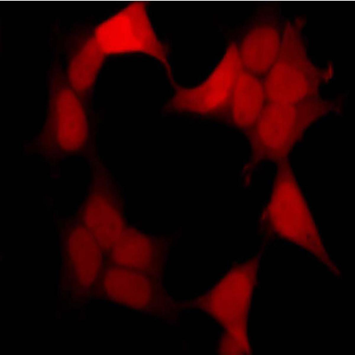
Immunofluorescent staining with anti GFP antibody, 1A5 |
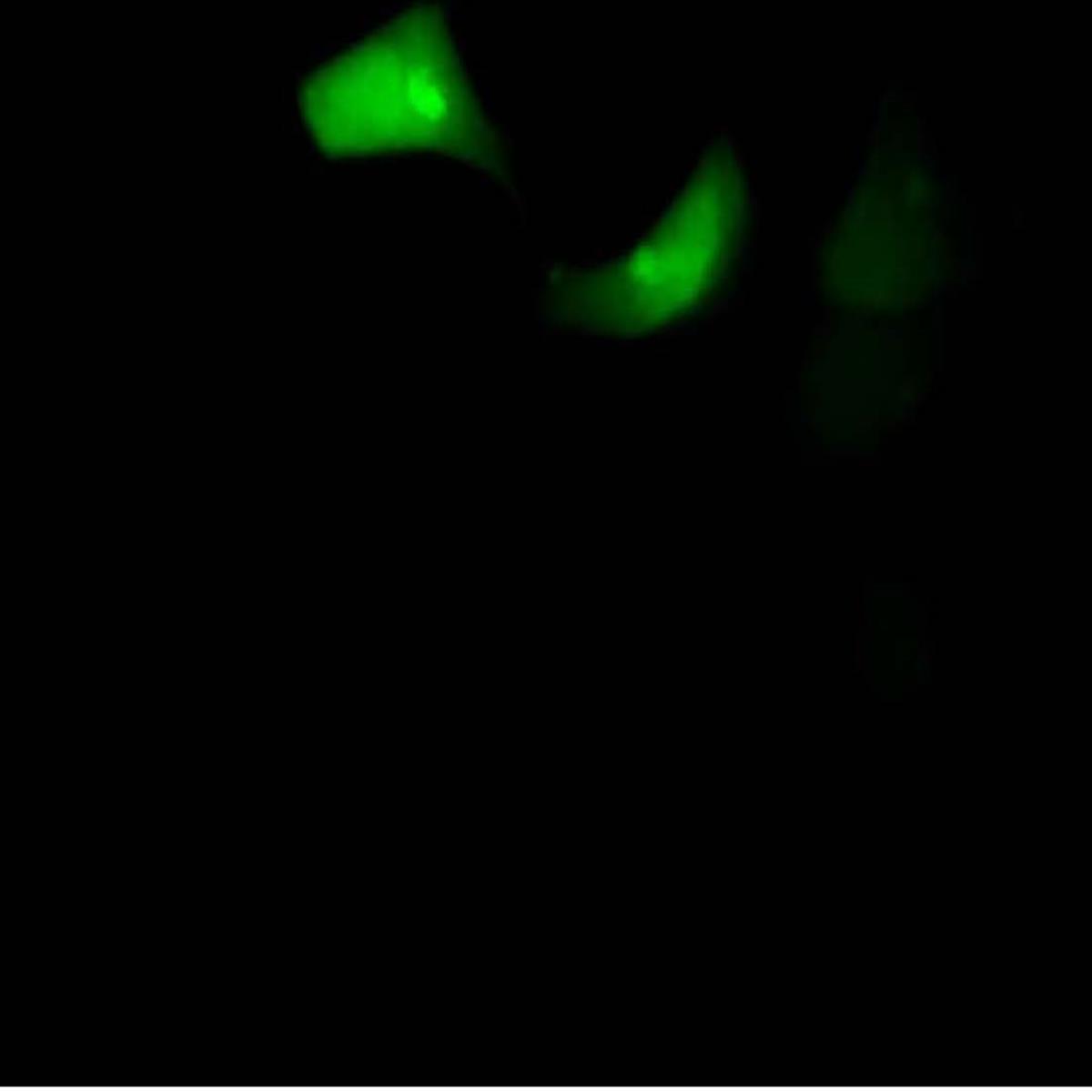
Fluorescent image due to GFP |


Fig.1 Fluorescent image of COS1 cells due to GFP of GST-ZIPK fusion protein expressed in HEK293T cells (Right) and the sams cells were immunostained by using anti-GFP antibody 1A5, followed by Texas Red-conjugated anti-rat IgG (Left). Note that fluorescence by the immunofluorescent staining using 1A5 antibody is much stronger than fluorescence due to GFP
|
|
|
|
|
Fig.3 Immunoprecipitation of GST-GFP fusion |


References
- Shimomura O et al (1962) "Extraction, purification and properties of aequorin, a bioluminescent protein from the luminous hydromedusan, Aequorea" J Cell Comp Physiol 59: 223–39 PMID: 13911999
- Chalfie M et al (1994) "Green fluorescent protein as a marker for gene expression" Science 263 (5148): 802–5 PMID: 8303295
- Tsien R (1998) "The green fluorescent protein" (PDF) Annu Rev Biochem 67: 509–44 PMID: 9759496