SGNPs#2
SGNPs#2<Sugar-immobilized Gold Nano-Particles#2>
Cat. No. FNK-G-AB2-250
Store at RT
Size 1set
Description
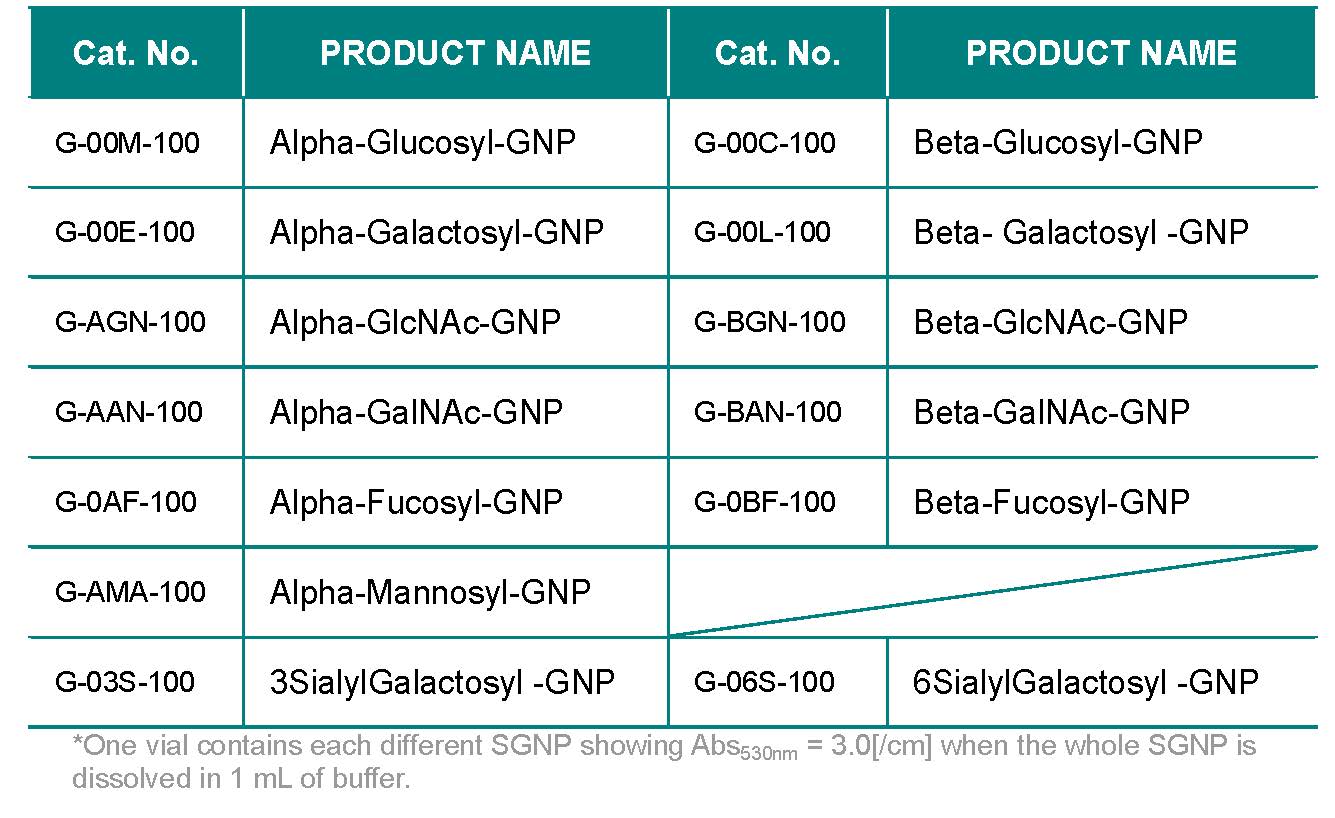
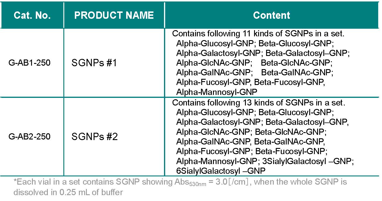
Kit Contains following 13 kinds of SGNPs in a set : α-Glucosyl-GNP, β-Glucosyl-GNP, α-Galactosyl-GNP, β-Galactosyl-GNP, α-GlcNAc-GNP, β-GlcNAc-GNP, α-GalNAc-GNP, β-GalNAc-GNP, α-Fucosyl-GNP, β-Fucosyl-GNP, α-Mannosyl-GNP, 3SialylGalactosyl-GNP, 6SialylGalactosyl-GNP
SGNP(Sugar chain - immobilized Gold Nano-Particle) is a gold nano particles immobilized with structurally defined sugar chains, showing red-purple color (λmax = ca. 530 nm). SGNP is a very convenient tool for evaluating sugar chain – protein interaction, since the interaction can be detected visually, and can be quantified from the change of OD at 530 nm. There are several applications as follows:
- Aggregation of protein
- Inhibition assay
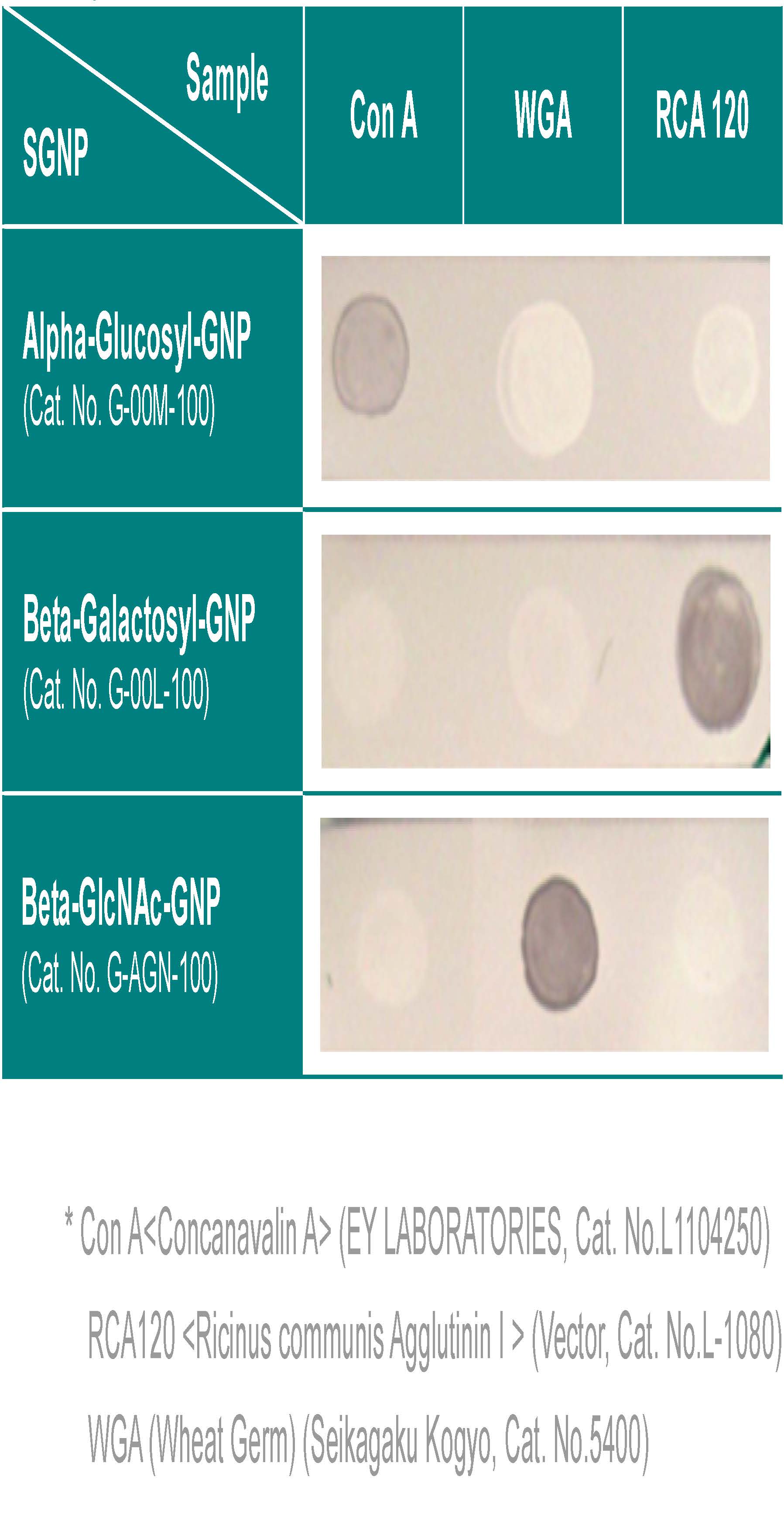
- Dot-blotting
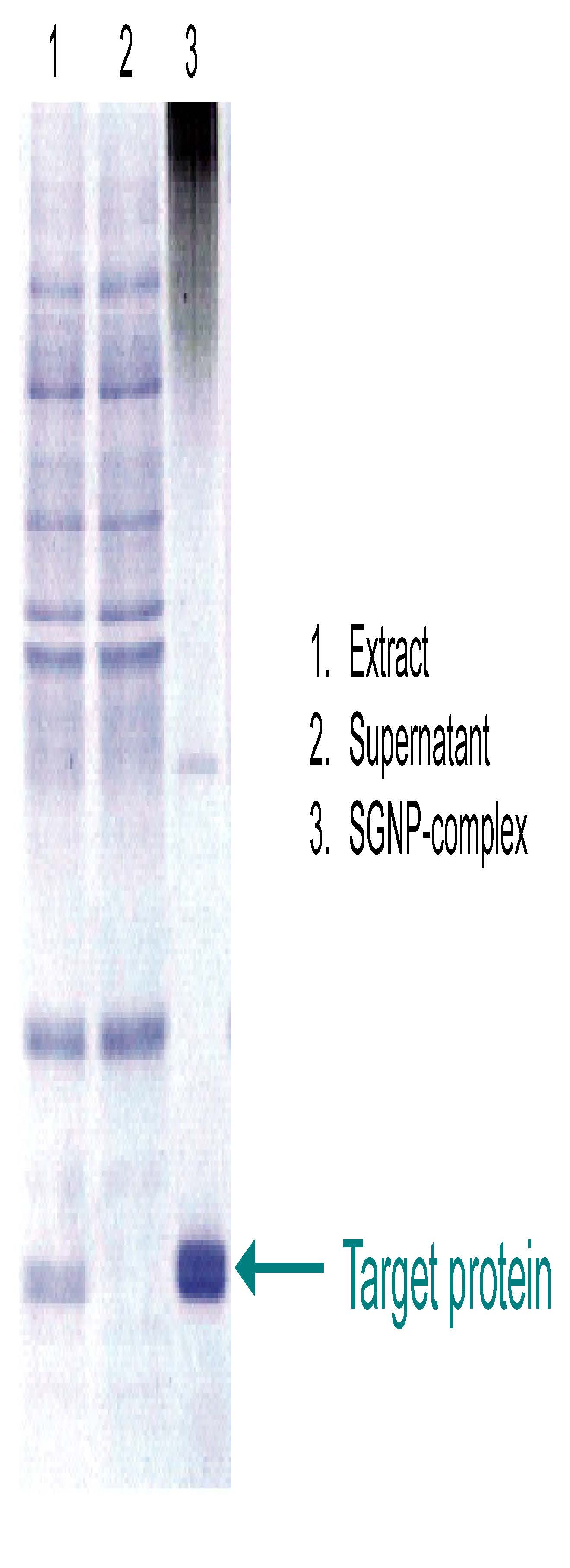
- Isolation and purification of lectin from a crude extract
SGNPs
SGNP set
Delivery as
Stability
Amount of SGNP in a vial
Abs530nm = 3.0[/cm] (dissolved in 1 mL of buffer)
Cat. No. G-***-250
Abs530nm = 3.0[/cm] (dissolved in 0.25 mL of buffer)
Addition of a reducing agent, such as mercaptoethanol, may affect the properties of SGNP.
Application #1: Aggregation assay of protein(s)
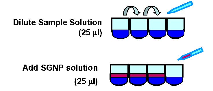
Example
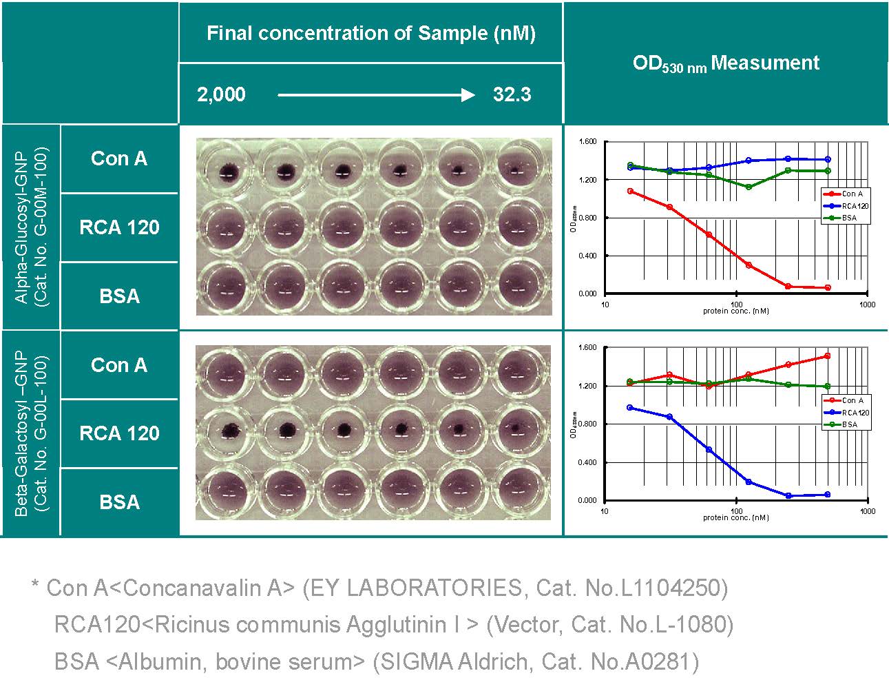
- Dissolved SGNP at Abs530nm = 3.0 using your buffer.
- *see Amount of SGNP
- In wells of 96-well microtiter plate (round bottom), 25μl of protein solution was added with changing the concentration.
- *Sequential 1:2 dilution from ca. 200 μg/ml (or ca. 4 μM), 6 points or more.
- Addition of 25 μl of SGNP solution prepared as above. Gentle agitation for 0.5 to 2 hr at room temperature.
- Measure OD at 530 nm of the supernatant.
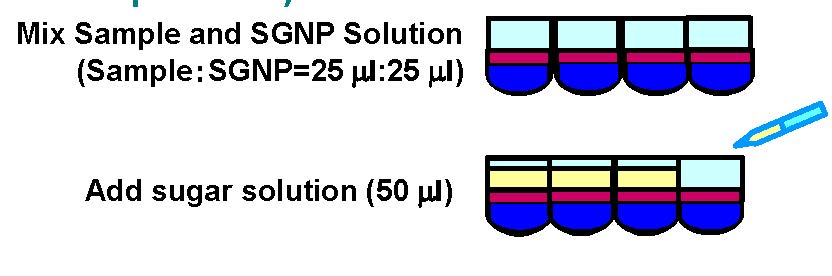
- Dissolve SGNP at Abs530nm = 4.0 ~ 6.0 using your buffer *see Amount of SGNP
- In wells of 96-well microtiter plate (round bottom), 25μl of protein solution (Conc. > KD) and 25 μl of SGNP solution as prepared above were added. *It is important to know KD value of the protein against sugar chain on SGNP
- 50 μl of the inhibitor dissolved in the same buffer (conc. 0.1 ~ 50 mM) is added to the above mixture. Then, agitate for 1 hr or overnight.
- Measure OD at 530 nm of the supernatant.
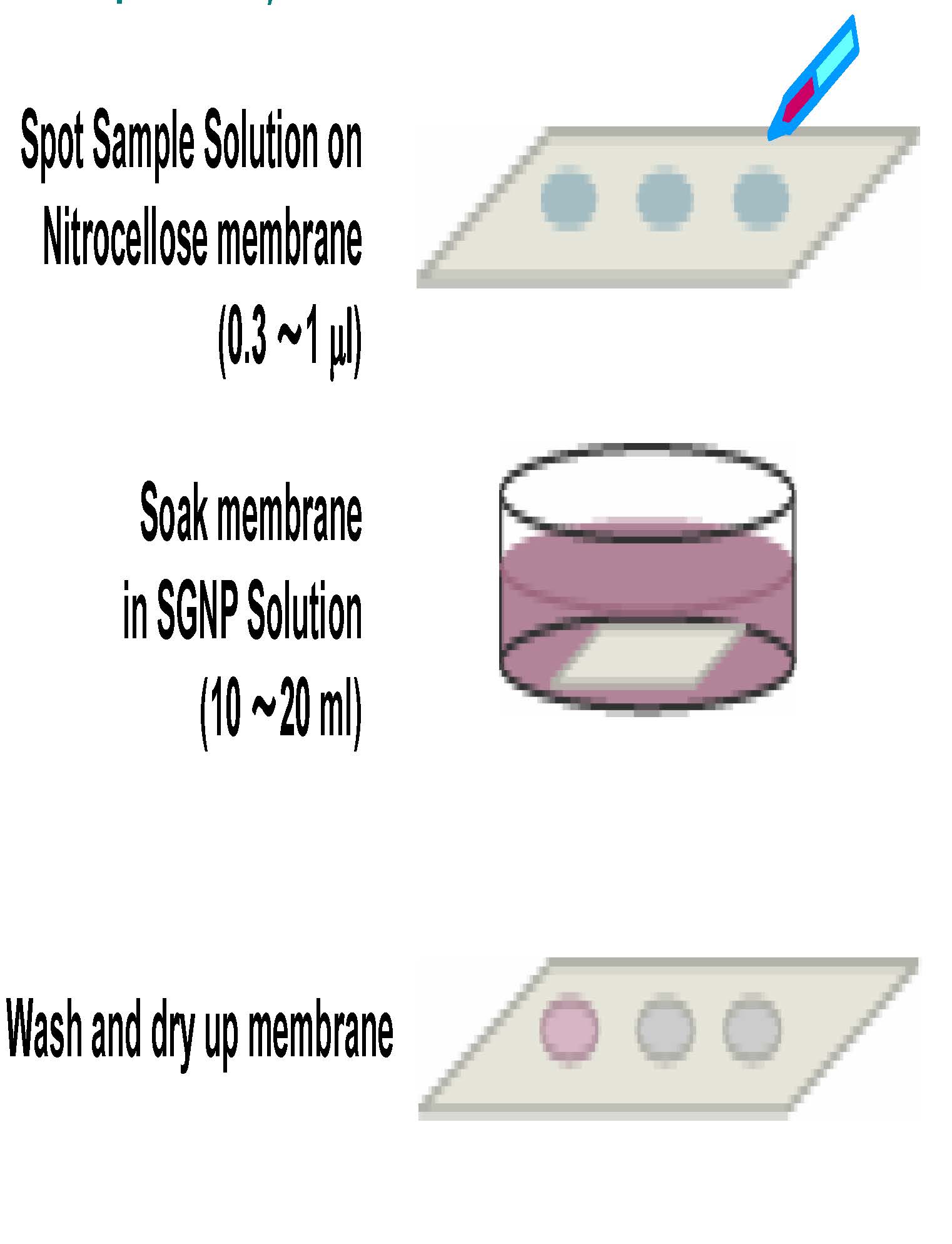
- Prepare 10~20ml SGNP solution of Abs530nm = 0.15 ~ 0.30 using your buffer. *see Amount of SGNP
- On nitrocellulose membrane* (10 x 30 mm), spot your sample (0.3 – 1 μL, 0.1- 2.0 μg) and dry up at room temperature. *Trans-BlotTM Transfer Medium, Pure Nitrocellose Membrane (0.2 μm) (Bio-Rad, Cat. No.162-0146)
- To 10 mL of SGNP solution in a glassware (φ 50 mm), the membrane is soaked with a gentle agitation for 5 – 30 min. *You may need to check the staining every 5 min. Please anchor the membrane with tweezers
- Wash the membrane with buffer and dry up. *If you see too high background, decrease the concentration of SGNP or shorten the soaking time.
directly applied for SDS-PAGE. Then, the protein band(s) in SDS-gel is analyzed according to the proteomics procedure.
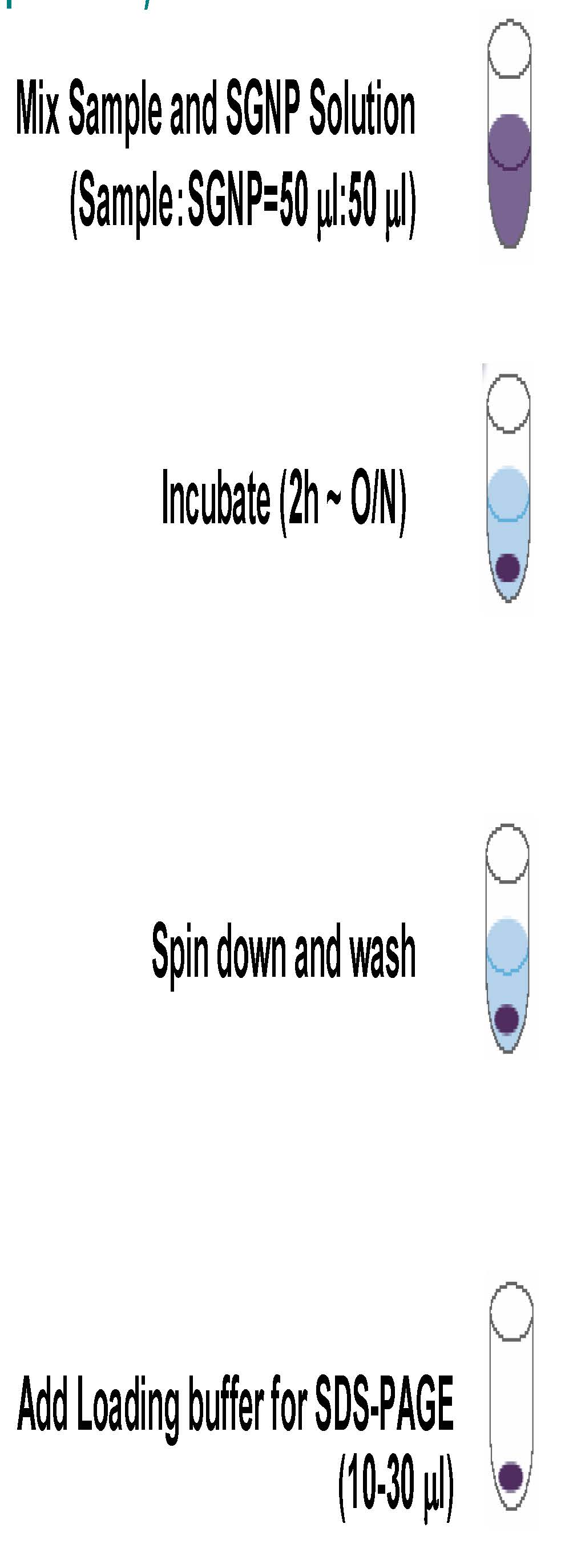
- Dissolve SGNP at Abs530nm = 3.0 using your buffer *see Amount of SGNP
- To 50 μl of SGNP solution in a 1.5 mL of eppendorf tube, add 50 μl of your extract. The mixture is incubated for 1 h to overnight at 4 degree C with a gentle agitation. * The protein concentration in the extract may be in the range between 500 and 10 mg/mL.
- Centrifugation at 6,000 ~ 10,000 x g for 10 min, and remove the supernatant.
- To the precipitate, add 500 μl of buffer, vortex for 10 sec and centrifuge at 6,000 ~ 10,000 x g for 10 min, and remove the supernatant. This may repeat 2 more times.
- To the precipitate, add 10-30 μl of sample preparation buffer*, boiled up for 10 min.. *Laemmli Sample Buffer (Bio-Rad, Cat. No.161-0737)
- SDS-PAGE *Reducing or non-reducing condition can be used.