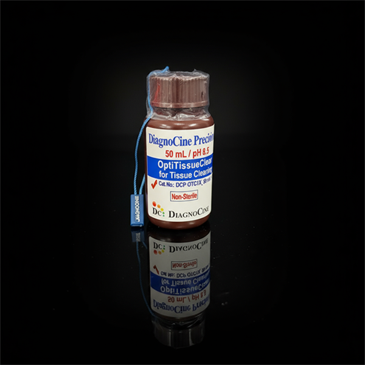
OptiTissueClear for Tissue Clearing
FluxMPS™ OptiTissueClear for Tissue Clearing
FluxMPS™ OptiTissueClear (Cat. No. DCP-OTC1X, 100 mL) is a next-generation tissue clearing reagent that delivers unparalleled optical transparency, superior fluorescence preservation, and accelerated clearing times for 3D microscopy applications — passing through a 0.22 µm membrane filter in a controlled manufacturing environment for reduced particulate burden.
- Multi-mechanism clearing: Combines lipid removal via micellar solubilization (SDS), hyperhydration (urea), penetration enhancement (DMSO), and antioxidant browning prevention (α-thioglycerol).
- Enhanced fluorescence: pH 8.5 alkaline borate buffer prevents fluorophore quenching and actively enhances signal intensity for GFP, RFP, and YFP.
- Deep antibody penetration: Synergistic combination of SDS, DMSO, and Tween 20 creates a permeable tissue matrix for faster antibody diffusion.
- Complete browning prevention: α-thioglycerol eliminates yellowing and autofluorescence throughout extended clearing procedures.
- Broad tissue compatibility: Compatible with most common fixation methods including 4% paraformaldehyde.
- 0.22 µm single-stage filtration: Filtered once through a 0.22 µm membrane in a non-sterile controlled environment.
- Includes RI-matched mounting solution: Refractive index = 1.52 for high-resolution confocal and light-sheet microscopy.
- ISO 13485 manufacturing: CE-approved facilities; assembled at DiagnoCine Precision, Totowa, New Jersey, USA.
- FormulationSDS + Urea + DMSO + α-thioglycerol + Tween 20 + Borate buffer
- pH (at 1X)8.5
- AppearanceClear solution
- Filtration0.22 µm membrane, once
- SterilityNon-sterile environment fill
- Clearing temperature42–47°C with gentle agitation
- Mounting solution RI1.52 (included)
- Storage2–8°C, away from bright light
- Shelf Life (unopened)12 months
- Shelf Life (after opening)6 months
Engineered where standard clearing protocols fall short
Conventional tissue clearing methods often compromise fluorescence signals, require days-long incubations, suffer from uneven clearing and browning artifacts, or use single-mechanism approaches that fail on dense tissues. FluxMPS™ OptiTissueClear combines four independent clearing mechanisms in a single, pH-controlled formulation that delivers faster clearing than standard CLARITY protocols while enhancing — not degrading — fluorescence signals.[1]
0.22 µm Filtered Purity
Single-pass 0.22 µm membrane filtration removes particulates that would introduce optical artifacts or interfere with refractive index uniformity during deep-tissue imaging.
Maximum Optical Clarity
Reduces light scattering through strategic lipid removal (SDS micellar solubilization) and refractive index matching, enabling high-resolution imaging deep within intact tissue volumes.[2]
Fluorescence Enhancement at pH 8.5
The alkaline borate buffer (pH 8.5) prevents fluorophore quenching and actively enhances signal intensity, preserving GFP, RFP, and YFP signals through extended clearing procedures.
Deep Antibody Penetration
Synergistic combination of SDS, DMSO, and Tween 20 creates a permeable tissue matrix that allows antibodies to penetrate faster and more uniformly into dense tissue specimens.[3]
Complete Browning Prevention
α-thioglycerol eliminates the yellowing and autofluorescence caused by Maillard reactions and oxidative damage that plague many clearing protocols, maintaining pristine optical properties throughout.
Customization on Demand
Other concentrations, pH values, chemical additions, or modified formulations are available. Contact support@diagnocine.com with your requirements.
Single-stage 0.22 µm filtration system
OptiTissueClear is filtered through a 0.22-micron membrane, once, in a controlled non-sterile manufacturing environment under ISO 13485:2016 quality management standards. This single-pass filtration removes large particulates and aggregates that would otherwise compromise optical clarity, refractive index uniformity, and imaging performance in three-dimensional microscopy workflows.
-
1
0.22 µmSingle-Pass Membrane Filtration
OptiTissueClear is passed through a 0.22 µm membrane filter exactly once prior to packaging. This removes particulate matter, aggregates, and undissolved components that could introduce optical artifacts during tissue clearing or interfere with refractive index matching. The filtration is performed in a controlled non-sterile manufacturing environment ? optimized for purity and clarity performance, not terminal sterilization.
Performance vs. unfiltered clearing reagents
Particulate-containing clearing reagents introduce light-scattering artifacts that degrade imaging depth and spatial resolution. By filtering at 0.22 µm, OptiTissueClear provides a consistent, particle-free baseline that ensures refractive index matching is not disrupted by suspended solids.
pore size
stage
© Diagnocine® — DCP-OTC1X
3D microscopy applications for complex tissue biology
OptiTissueClear is engineered for researchers requiring reliable optical clearing across a wide range of tissue types, fixation methods, and fluorescent reporters. The multi-mechanism formulation supports whole-organ imaging, deep-tissue immunofluorescence, and advanced 3D reconstruction workflows.
Microfluidic & Chip-Compatible Use
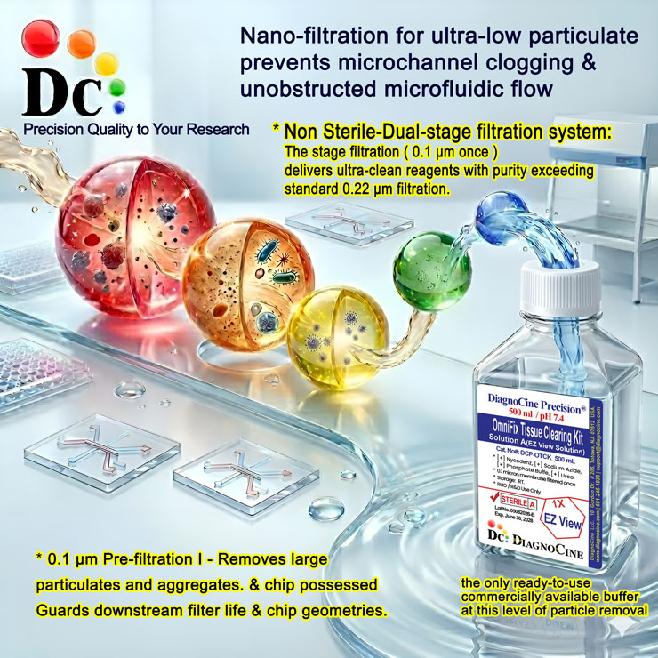
The 0.22 µm filtered OptiTissueClear reagent is compatible with microfluidic tissue-on-a-chip (ToC) and organ-on-a-chip (OoC) platforms for on-chip tissue clearing workflows. For automated bioreactor and robotics applications requiring the highest purity standard, a custom ultra-filtered variant is available on inquiry.
- Reduced Channel Contamination: 0.22 µm filtration minimizes particulate load in microfluidic clearing workflows, reducing clogging risk.
- Consistent RI Matching: Particle-free formulation ensures reproducible refractive index uniformity across chip-scale tissue preparations.
- Thermal Compatibility: Clearing at 42–47°C is compatible with most microfluidic platform thermal management systems.
Inquiry Required: Custom purity grades and formulation modifications are available on request. Contact support@diagnocine.com with your system specifications.
3D Reconstruction & Anatomy
Delivers the optical transparency required for whole-organ imaging and anatomical phenotyping. Compatible with light-sheet fluorescence microscopy (LSFM) for intact specimen reconstruction.[4]
Neural Circuit Tracing & Connectomics
Enables intact-brain imaging for connectome analysis. Preserves fluorescent protein expression (GFP, RFP, YFP) for tracing axonal projections and mapping synaptic connectivity across brain regions.[5]
Tumor Microenvironment Analysis
Clears dense tumor specimens to reveal immune cell infiltration, vasculature architecture, and stromal organization in 3D. Compatible with multiplex immunofluorescence staining protocols.
Embryo & Developmental Studies
Gentle pH-controlled formulation preserves tissue morphology for developmental staging studies, organ primordium analysis, and lineage tracing with fluorescent reporters across developmental time points.
Deep-Tissue Immunofluorescence
SDS + DMSO + Tween 20 combination creates a permeable tissue matrix enabling antibody penetration into thick sections (1–5 mm). Supports primary/secondary antibody staining after clearing.[3]
Multi-Color Fluorescent Protein Imaging
Compatible with GFP, RFP, YFP and most common fluorescent proteins. pH 8.5 formulation actively enhances fluorescence signal intensity rather than quenching it during extended clearing incubations.
Analytical & quality specifications
All parameters measured per lot and documented in the Certificate of Analysis. Contact support@diagnocine.com to request a CoA for a specific lot number.
| Parameter | Specification |
|---|---|
| Formulation | SDS, Urea, DMSO, α-thioglycerol, Tween 20, Borate buffer |
| Appearance | Clear solution |
| pH USP <791> | 8.5 (at 1X concentration) |
| Buffer system | Alkaline borate buffer |
| Refractive index matching | Mounting solution RI = 1.52 (included) |
| Clearing temperature | 42–47°C with gentle agitation |
| Parameter | Specification |
|---|---|
| Filtration pore size | 0.22 µm membrane |
| Filtration stages | 1 (single-pass) |
| Fill environment | Non-sterile controlled manufacturing environment |
| Sterility declaration | Non-sterile; filtered 0.22-micron membrane once |
| Manufacturing ISO 13485 | ISO 13485:2016 QMS; CE-approved supplier |
| pH monitoring (use) | Replace if pH drops below 8.0 during clearing |
| Parameter | Specification |
|---|---|
| Storage temperature | 2–8°C, away from bright light |
| Pre-use preparation | Must warm to room temperature before use |
| Shelf life (unopened) | 12 months |
| Shelf life (after opening) | 6 months |
| Expiry date | Use before expiry date on product label |
| Solution exchange | Replace every 24–48 h during clearing for consistency |
| Parameter | Specification |
|---|---|
| Raw material grade | Research/laboratory grade; lot-traceable |
| Manufacturing QMS ISO 13485 | ISO 13485:2016 certified |
| Regulatory alignment | CE-approved supplier |
| Production site | DiagnoCine Precision, Totowa, New Jersey, USA |
| Intended use | For Research Use Only (RUO) |
| Safety classification | Contains hazardous chemicals; see Safety section |
Full composition
OptiTissueClear contains a proprietary multi-component formulation combining four independent clearing mechanisms. Exact concentrations are proprietary. CAS numbers are provided for informational and safety reference. DMSO is a skin-penetrating solvent — always handle with appropriate chemical-resistant gloves (see Safety section below).
| Ingredient | CAS Number | Role / Concentration |
|---|---|---|
| CLEARING AGENTS | ||
| Sodium Dodecyl Sulfate (SDS) | 151-21-3 | Lipid removal via micellar solubilization; proprietary concentration |
| Urea | 57-13-6 | Hyperhydration; prevents tissue shrinkage and reduces light scattering; proprietary concentration |
| Dimethyl Sulfoxide (DMSO) | 67-68-5 | Penetration enhancer; accelerates diffusion into dense tissues; proprietary concentration |
| Tween 20 (Polysorbate 20) | 9005-64-5 | Surfactant; contributes to permeable tissue matrix for antibody penetration; proprietary concentration |
| Ingredient | CAS Number | Role / Concentration |
|---|---|---|
| BUFFER & ADDITIVES | ||
| Borate buffer (alkaline) | 10043-35-3 (boric acid component) | pH control at 8.5; prevents fluorophore quenching; proprietary concentration |
| α-thioglycerol | 96-27-5 | Antioxidant; eliminates browning (Maillard reactions) and autofluorescence; proprietary concentration |
| Antioxidants (proprietary blend) | Proprietary | Mitigates oxidative damage to fluorophores and tissue proteins; proprietary concentration |
| Parameter | Compatibility | Notes |
|---|---|---|
| FIXATION & FLUORESCENT PROTEIN COMPATIBILITY | ||
| 4% Paraformaldehyde (PFA) | Primary validated fixation | Fix per standard protocols before clearing |
| Most common fixation methods | Compatible | Validate for each tissue type; monitor pH during clearing |
| GFP (Green Fluorescent Protein) | Preserved | pH 8.5 buffer actively enhances GFP signal |
| RFP (Red Fluorescent Protein) | Preserved | Antioxidant protection prevents signal degradation |
| YFP (Yellow Fluorescent Protein) | Preserved | Alkaline pH maintains YFP fluorescence throughout clearing |
| Antibody immunostaining (post-clearing) | Compatible | SDS + DMSO + Tween 20 enhance deep antibody penetration |
OptiTissueClear protocol & usage guidelines
Follow the three-stage workflow below for optimal tissue clearing results. Monitor pH daily during the clearing process and tailor time and temperature to tissue size and lipid content. Replace clearing solution every 24–48 hours for consistency.
Tissue Preparation
Fix tissue samples in 4% paraformaldehyde according to standard protocols. OptiTissueClear is compatible with most common fixation methods and fluorescent proteins including GFP, RFP, and YFP.
Clearing Process
Immerse fixed tissue in OptiTissueClear solution at 42–47°C with gentle agitation. Replace solution every 24–48 hours for optimal results and monitor pH daily — replace if pH drops below 8.0. Clearing time varies by tissue type: soft tissues (spleen, brain) clear faster while dense tissues (stomach, bone-containing samples) require extended incubation. Scientists should optimize clearing duration for their specific tissue of interest.
Mounting for Imaging
After achieving desired transparency, transfer tissue to the included refractive index-matched mounting solution (RI = 1.52) for 24–48 hours at 37°C. This final step eliminates residual refractive index mismatches and prepares samples for high-resolution confocal or light-sheet microscopy.
warningSafety & Handling — Hazardous Chemical Warning
For Research Use Only. OptiTissueClear contains hazardous chemicals including corrosive detergents, skin-penetrating solvents (DMSO), and reproductive toxicants. Always wear:
- Chemical safety goggles
- Chemical-resistant gloves — butyl rubber, neoprene, or thick nitrile ≥15 mil (never use thin nitrile gloves with DMSO)
- Laboratory coat
Work in a properly ventilated area or fume hood, especially when opening containers or changing solutions. In case of eye contact, immediately flush with water for 15–20 minutes and seek medical attention. For skin contact, wash thoroughly with soap and water for at least 15 minutes. Never pour used solution down the drain — dispose of all waste as hazardous chemical waste through your institution's Environmental Health & Safety department. Pregnant personnel should consult occupational health before handling due to reproductive hazard components. Keep away from strong oxidizers and acids.
Manufacturing standards & compliance
OptiTissueClear is manufactured under ISO 13485-certified and CE-approved facility standards. All final packaging, quality assurance, and testing are performed at the DiagnoCine R&D and Quality Testing Center. All specific customization requests and assembly are accomplished at DiagnoCine Precision in Totowa, New Jersey, USA.
ISO 13485:2016 QMS
Manufactured under a full ISO 13485:2016 quality management system with CE-approved supplier status. Products released only after QC testing at DiagnoCine R&D and Quality Testing Center.
0.22 µm Single-Pass Filtration
Every production batch is filtered through a 0.22 µm membrane (single pass) in a controlled non-sterile manufacturing environment, removing particulates that would compromise optical clarity and RI matching.
DiagnoCine Precision Assembly
All final kit assembly, labeling, customization, and quality release performed at DiagnoCine Precision, Totowa, New Jersey, USA under controlled laboratory conditions.
Lot-Specific CoA
Each lot is accompanied by a Certificate of Analysis documenting pH (8.5), appearance, filtration grade, and shelf-life validation. Contact support@diagnocine.com for lot-specific CoA.
pH Verification
pH 8.5 at 1X concentration measured per lot by calibrated pH meter. Product must be replaced during use if pH drops below 8.0; documented in CoA.
Appearance QC
Clear solution on incoming and outgoing inspection. Any turbidity or particulate contamination constitutes a lot-failure event requiring investigation and reprocessing.
Filtration Verification
0.22 µm membrane filtration applied to every batch. Filter integrity tested before and after filtration per internal protocol aligned with ISO 13485 requirements.
Documentation & Traceability
Full batch records including raw material traceability, filtration logs, QC results, and CoA maintained. Hazardous component documentation available for institutional compliance upon request.
How DCP-OTC1X compares to standard clearing approaches
OptiTissueClear benchmarked against commonly used single-mechanism clearing protocols to highlight advantages in speed, fluorescence preservation, and browning prevention.
| Parameter | DCP-OTC1X (FluxMPS™ OptiTissueClear) | Standard CLARITY Protocol | Generic SDS Clearing (0.22 µm) |
|---|---|---|---|
| Clearing mechanism | Multi-mechanism (SDS + Urea + DMSO + α-thioglycerol) | Single-mechanism SDS electrophoresis | Single-mechanism SDS passive |
| Filtration pore size | 0.22 µm single-pass | Typically unfiltered | 0.22 µm (when filtered) |
| Fluorescence preservation | check_circle Enhanced at pH 8.5 | cancel Quenching risk | cancel Variable; pH-dependent |
| Browning prevention | check_circle α-thioglycerol included | cancel No specific protection | cancel Not addressed |
| Antibody penetration | check_circle Enhanced (SDS+DMSO+Tween 20) | check_circle Moderate | cancel Limited |
| Ready-to-use format | check_circle Yes | cancel Manual preparation required | cancel Manual preparation |
| RI mounting solution | check_circle Included (RI = 1.52) | cancel Separate purchase | cancel Separate purchase |
| Manufacturing QMS | ISO 13485:2016; CE-approved | No certification | Varies by supplier |
| GFP/RFP/YFP compatible | check_circle Validated | check_circle Variable | cancel Not validated |
| Custom formulation | check_circle Available on request | cancel No | cancel No |
Frequently asked questions
Common questions about OptiTissueClear Tissue Clearing Reagent (DCP-OTC1X).
Supporting literature
Curated peer-reviewed references on SDS-based tissue clearing, fluorescence preservation, deep-tissue immunofluorescence, and advanced 3D microscopy of intact specimens.
- Chung K, et al. Structural and molecular interrogation of intact biological systems. Nature. 2013;497(7449):332–337.doi:10.1038/nature12107
- Susaki EA, et al. Whole-brain imaging with single-cell resolution using chemical cocktails and computational analysis. Cell. 2014;157(3):726–739.doi:10.1016/j.cell.2014.03.042
- Renier N, et al. iDISCO: a simple, rapid method to immunolabel large tissue samples for volume imaging. Cell. 2014;159(4):896–910.doi:10.1016/j.cell.2014.10.010
- Tomer R, et al. Advanced CLARITY for rapid and high-resolution imaging of intact tissues. Nature Protocols. 2014;9(7):1682–1697.doi:10.1038/nprot.2014.123
- Susaki EA, Ueda HR. Whole-body and whole-organ clearing and imaging techniques with single-cell resolution: toward organism-level systems biology in mammals. Cell Chemical Biology. 2016;23(1):137–157.doi:10.1016/j.chembiol.2015.11.009
- Pan C, et al. Shrinkage-mediated imaging of entire organs and organisms using uDISCO. Nature Methods. 2016;13(10):859–867.doi:10.1038/nmeth.3964
- Zhao S, et al. Cellular and molecular probing of intact human organs. Cell. 2020;180(4):796–812.doi:10.1016/j.cell.2020.01.030
- Ariel P. A beginner's guide to tissue clearing. International Journal of Biochemistry & Cell Biology. 2017;84:35–39.doi:10.1016/j.biocel.2016.12.009
- Marx V. Optimizing probes to image cleared tissue. Nature Methods. 2016;13(3):205–208.doi:10.1038/nmeth.3774
- Richardson DS, Lichtman JW. Clarifying tissue clearing. Cell. 2015;162(2):246–257.doi:10.1016/j.cell.2015.06.067