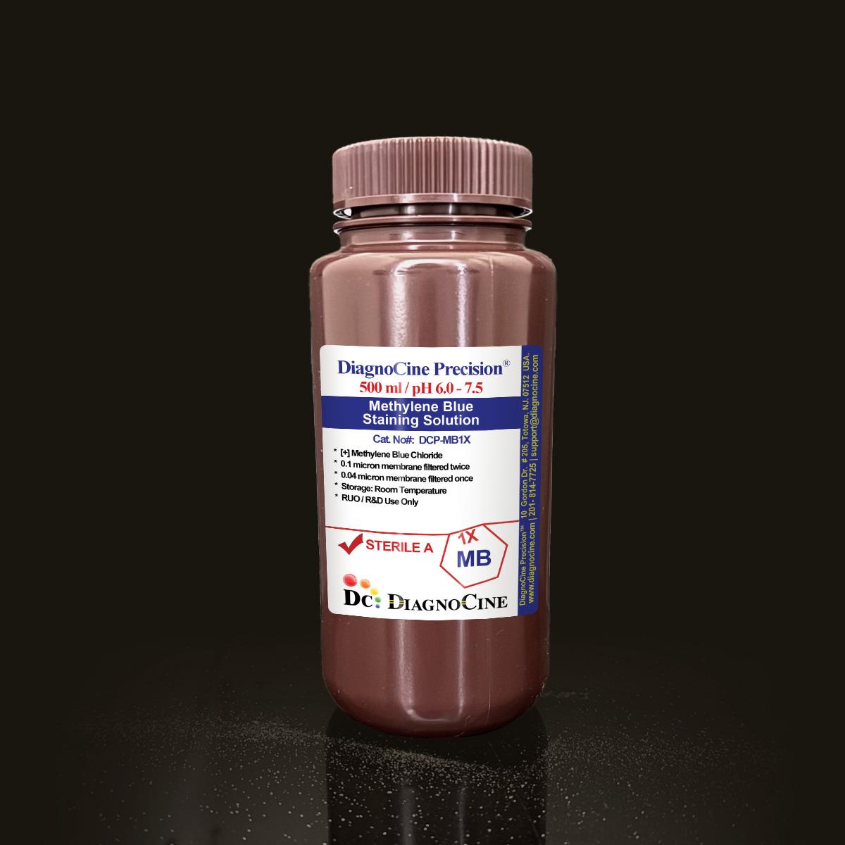
Methylene Blue Staining Solution
Cat No.: DCP-MB1X
Size: 500 mL
Sterile, Ultrapure, Filtered 0.1-micron membrane TWICE and 0.04-micron membrane ONCE.
Description:
Methylene Blue staining solution represents a standardized biological dye formulation optimized for diverse laboratory applications requiring precise cellular visualization and diagnostic staining. This ready-to-use aqueous solution combines the chemical stability of methylene blue with rigorous quality control measures to ensure consistent performance across microbiology, histology, and molecular biology workflows. Methylene Blue staining solution is a 1% (w/v) aqueous preparation of methylene blue chloride (C16H18ClN3S · xH₂O), a phenothiazine-derived cationic dye with a molecular weight of 319.85 g/mol. The solution appears as a deep blue liquid with a mild chemical odor, formulated to pH 6.0–7.5 for compatibility with biological specimens. Its positively charged molecules exhibit selective binding to negatively charged cellular components, enabling high-contrast visualization of nuclei, RNA-rich cytoplasmic regions, and bacterial morphology under light microscopy.
Key Features:
The solution demonstrates exceptional batch-to-batch consistency, with ≥99% purity of the active methylene blue component. Its low viscosity and water-like density (~1.00 g/cm³) facilitate easy pipetting and even specimen coverage during staining protocols. Unlike ethidium bromide-based stains, methylene blue does not intercalate nucleic acids, preserving DNA/RNA integrity for downstream hybridization processes while providing sufficient sensitivity for routine gel electrophoresis applications.
Applications:
Microbiology & Bacteriology
- Gram Staining: Serves as a counterstain in modified Gram protocols, differentiating Gram-negative bacteria through selective retention after decolorization.
- Capsule Visualization: Highlights polysaccharide capsules as unstained halos around blue-stained bacterial cells via negative staining techniques.
- Viability Assays: Distinguishes live/dead yeast and microbial cultures through redox activity—viable cells reduce the dye to colorless leukomethylene blue.
Histology & Cytology
- Nuclear Staining: Intensely stains nuclei dark blue while lightly coloring cytoplasm, ideal for peripheral blood smears and tissue sections.
- Special Stains: Optimized for Ziehl-Neelsen acid-fast bacilli (AFB) staining and Fite's modified AFB protocols for leprosy diagnostics.
Molecular Biology
- Nucleic Acid Detection: Visualizes RNA/DNA in northern/Southern blots and agarose gels without interfering with membrane hybridization.
- Metachromatic Analysis: Exhibits concentration-dependent spectral shifts (λmax 664 nm monomeric, 605 nm dimeric) for studying dye aggregation phenomena.
Composition:
| Ingredients |
Concentration |
| Methylene Blue Chloride |
1% |
Usage Instructions:
General Protocol for Microscopy Staining
-
Specimen Preparation:
-
For liquid samples: Apply 10–20 μL directly to a clean glass slide.
-
For solid tissues: Prepare thin sections (4–6 μm) using a microtome.
-
Air-dry specimens completely; heat-fix bacterial smears by briefly passing through a flame (except for capsule staining).
-
Staining:
-
Post-Staining:
-
Microscopy:
Safety Information:
- Protective Measures: Nitrile gloves, lab coat, and eye protection required.
- Spill Management: Absorb with vermiculite; dispose as hazardous waste.
- Incompatibilities: Strong oxidizers, acids.
- Decontamination: Clean equipment with 70% ethanol followed by soap-water rinse.
Quality Control:
- Appearance: Dark blue solution
- pH: 6.0–7.5
- Sterility: Filtered 0.1 micron Twice and 0.04 micron once.
- DNase activity: None was detected after incubation of plasmid DNA, and this product was used for 18 hours at room temperature.
- RNase activity: No RNase activity was detected after incubation of ribosomal RNA and this product for 18 hr at room temperature.
Storage and Shelf Life:
- Store at room temperature away from bright light.
- Shelf life is 24 months.
- Use before expiry date given on the product label.
CUSTOMIZATION: Please INQUIRE if other concentrations, additions of chemicals/compounds/proteins/supplements, different pH, and modifications are needed.
All DiagnoCine Precision Sterile buffers are filtered-sterilized with a 0.1-micron filtration two times and 0.04-micron filtration one time. Thus, mycoplasma contamination is prevented. The smallest size mycoplasma type can be about 0.2 microns.
* This product is manufactured under ISO 13485-certified and CE-approved facilities (Suppliers of DiagnoCine Precision). All final packaging, quality assurance, and testing are done at the DiagnoCine R&D and Quality Testing Center. All specific customization requests and assembly were accomplished at DiagnoCine Precision in Totowa, New Jersey, USA.