FAOBlue (Fatty Acid Oxidation Detection Reagent)
FAOBlue (Fatty Acid Oxidation Detection Reagent)
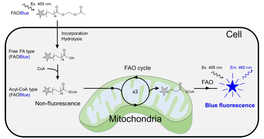
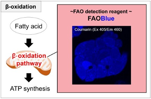
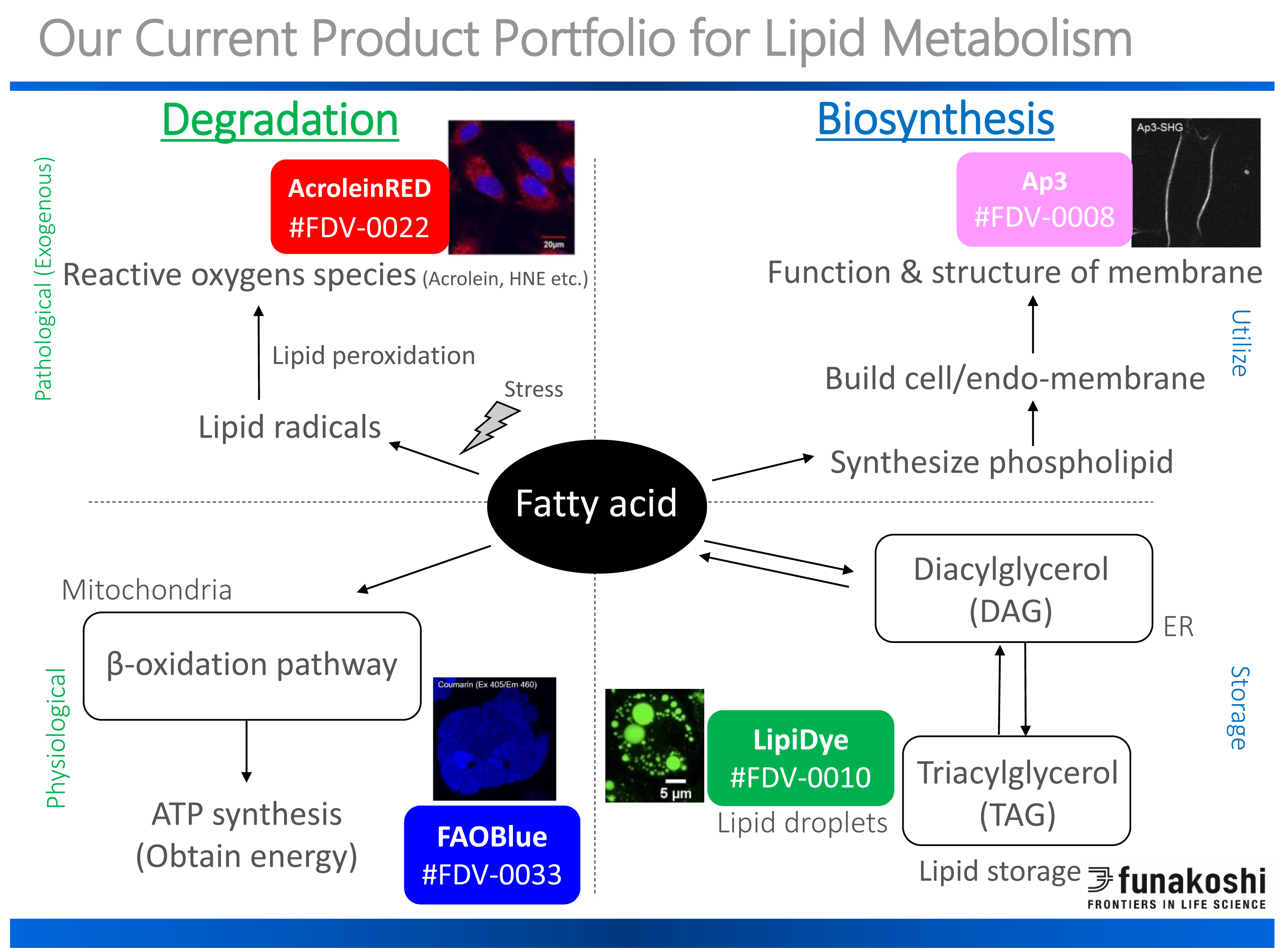
FAOBlue Directly Detect Fatty Acid Oxidation (FAO) Activity in LIVE CELLS. FAO Blue is a newly designed fluorescent detection system (Excitation 405nm: Emission 460nm) to study catabolic pathway for energy production. Mitochondria and associated enzymes in the catabolic pathways can be detected to measure FAO activities by simply adding the dye to your cell.
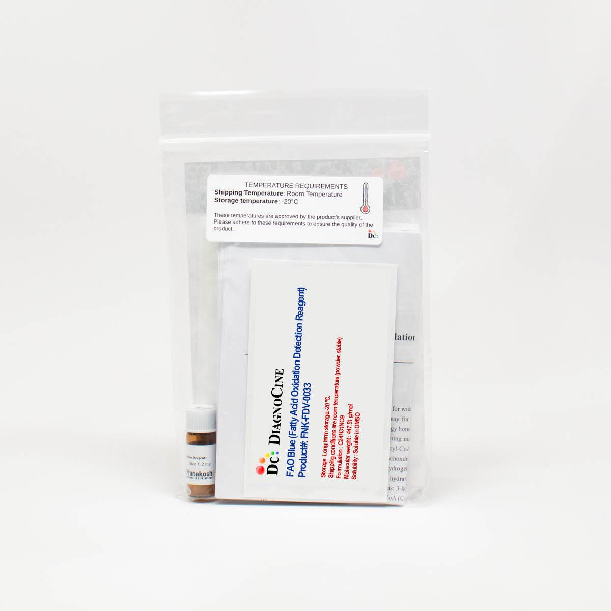
Cat.No. FNK-FDV-0033
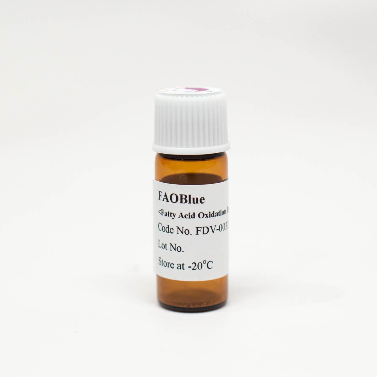
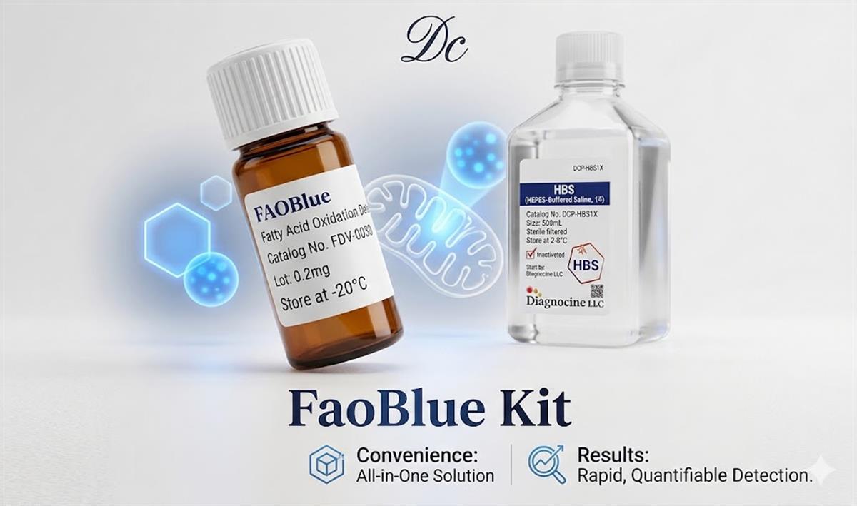
(a) FaoBlue Reagent
Size 0.2 mg
Storage Long term storage:-20 ºC. Shipping conditions are room temperature (powder, stable)
Formulation : C24H31NO9
Molecular weight : 447.51 g/mol
Solubility : Soluble in DMSO
(b) HEPES-buffered saline (HBS buffer)
Size 500ml
Sterile: Filtered 0.1-micron membrane TWICE, 0.04-micron membrane ONCE
| tip | The Best Match Bundle Products for Lipid Metabolism Research | LipiDye II, Lipid dye Droplet Staining with FAO Blue (Fatty Acid Oxidation Detection Reagent |  |
Description
* This product has been commercialized and sold by Funakoshi Co., Ltd. based on the research results of Professor Akio Ojida, a chemical biology field at the Graduate School of Pharmaceutical Sciences, Kyushu University.
* This product is for research. It can only be used for research.
 |
or, Go Webinar FAO Blue (Fatty Acid Oxidation Detection Reagent) >>> Click here <<<
Product Background
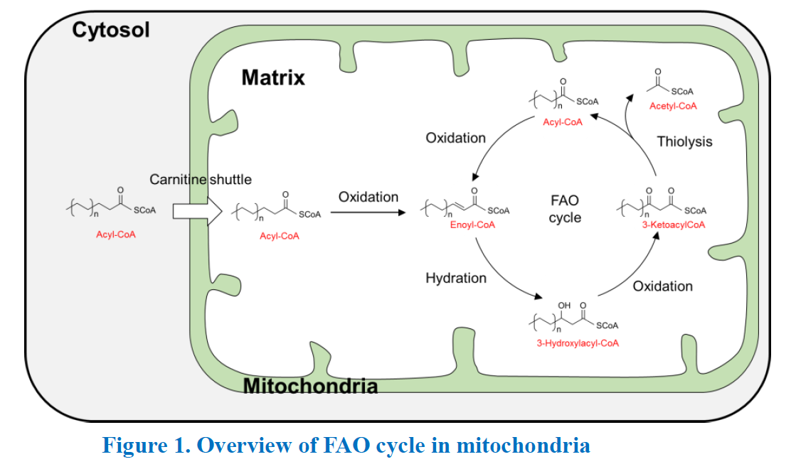
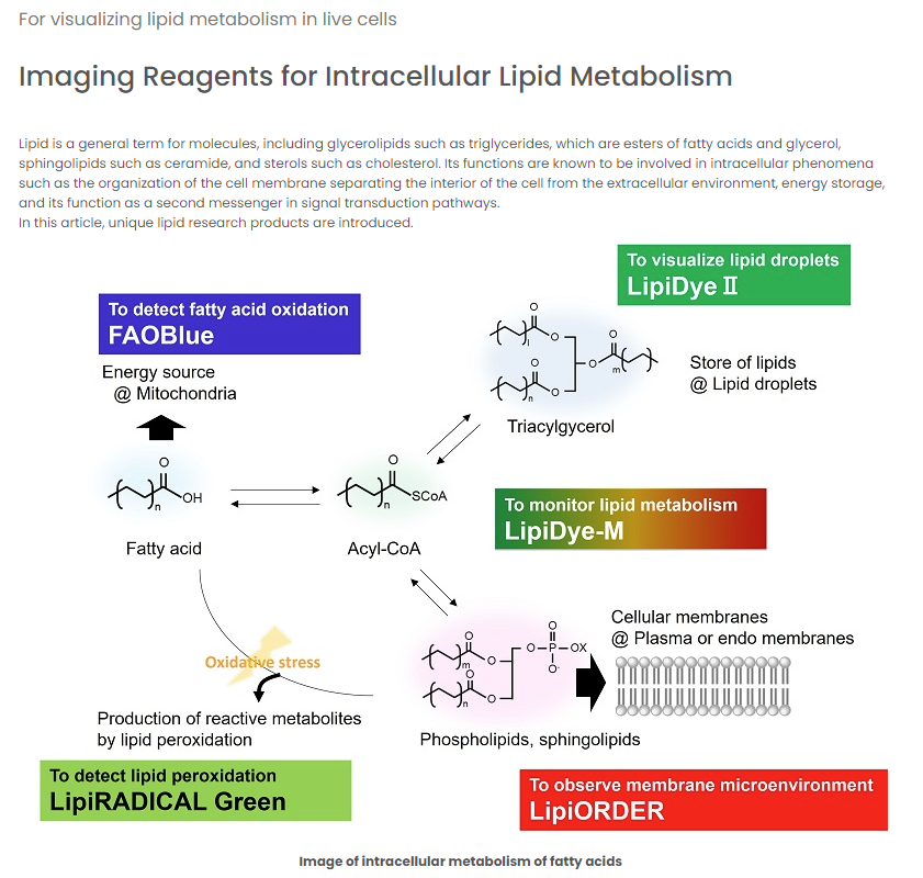
Fatty acids (FAs) are basic building blocks for wide variety of lipids, essential components of cells, and are one of primary sources of energy. The major pathway for the degradation of FAs is mitochondrial FA beta-oxidation (FAO). FAO is a key metabolic pathway for energy homeostasis in organs such as the liver, heart and skeletal muscle. FAO is a complicated biochemical event containing many types of enzymes. First, FAs are converted to acyl-CoA form by acyl-CoA synthetase family. Second, acyl-CoA forms are incorporated into mitochondria via the carnitine shuttle pathway. Once acyl-CoA entering to mitochondrial matrix, acyl-CoA (Cn) is converted to acyl-CoA (Cn-2) and acetyl-CoA by four stepwise reactions. 1) Dehydrogeneration: Acyl-CoA is oxidized to enoyl-CoA by acyl-CoA dehydrogenases, 2) Hydration: Enoyl-CoA is hydrated to 3-hydroxyacyl-CoA by crotonase, 3) Oxidation: 3-hydroxyacyl-CoA to 3-ketoacyl-CoA, 4) Thiolysis: 3-ketoacyl-CoA to acyl-CoA (Cn-2) and acetyl-CoA. Acetyl-CoA is further converted to ATP. The resulting acyl-CoA (Cn-2) enters another cycle of FAO to further produce acyl-CoA (Cn-4).
Abnormal FAO is involved in various diseases such as obesity and non-alcoholic fatty liver diseases (NAFLD). Although measurement of FAO activity in diseased cells is important, methods for measuring FAO activity are limited due to its complicated processes above. Only a few indirect methods such as using radio-isotope containing fatty acids or measuring oxygen consumption are commonly performed.
Application
- Relative quantification of FAO activity
- Evaluation of drug effect on FAO activity
Reconstitution and Storage
Reconstitution : Add 100% DMSO into vial to prepare 1-10 mM stock solution.
Storage :
Stock solution :
- Make aliquots and store at -20u°C with protection from light.
- Avoid repeated freeze-thaw cycles.
General procedure
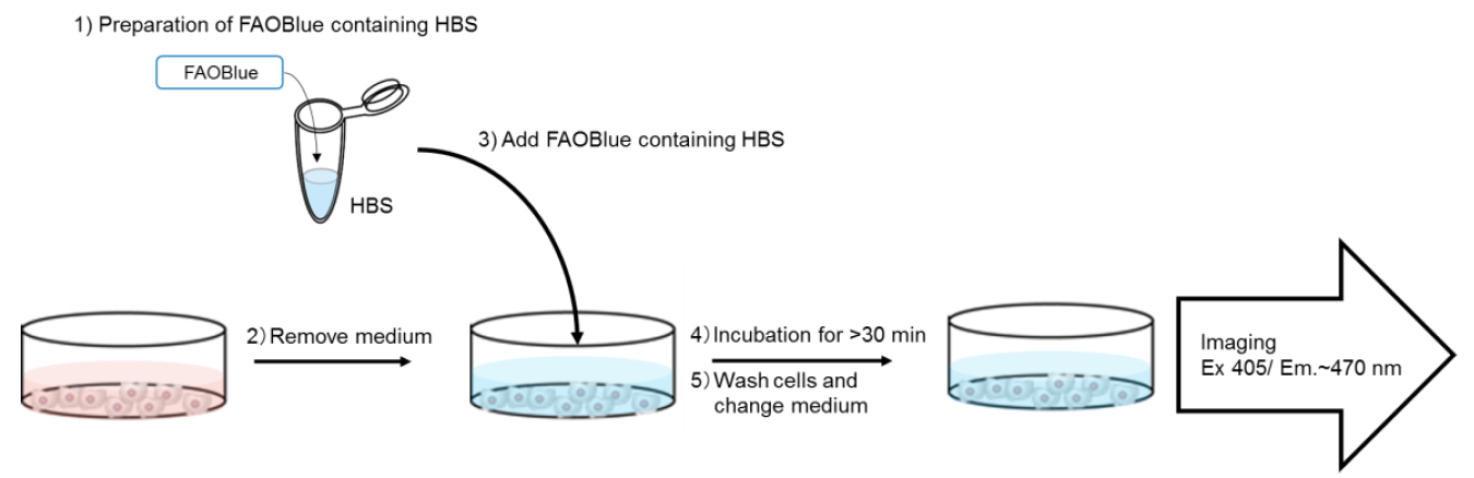
1. Add FAOBlue (recommended final conc. 5-20 ?M) in fresh HEPES-buffered saline (HBS)
3. Add FAOBlue-containing HBS to cells
4. Incubate cells at 37oC for >30 min
5. Wash cells with HBS
6. Observe cells under live condition with blue fluorescence (Ex 405 nm/ Em. 430-480 nm)
- Protocol (from Bio-protocol) can be found here [LINK]
- GO "Flow Cytometry" -- Click here --
Reference data and Experimental guide
| Absorbance and fluorescence spectrum of FAOBlue and coumarin-derivative |
|
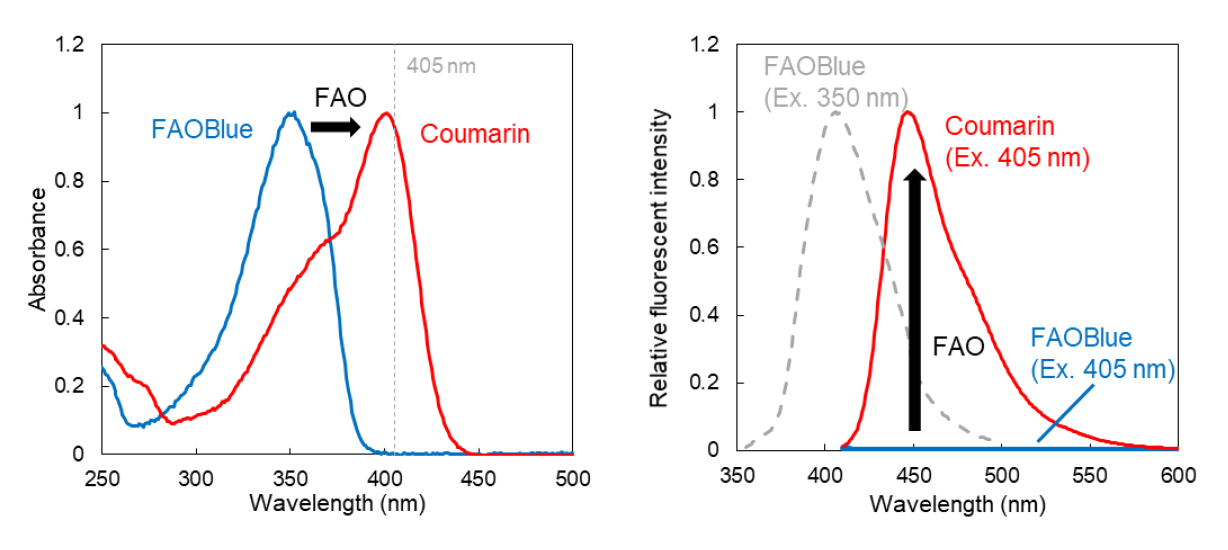
Absorbance (left) and fluorescence (right) spectra of FAOBlue (blue line) and the coumarin-derivative dye released after metabolization of FAO (red line) in PBS buffer (pH 7.4).
Absorbance spectra: an absorption peak of coumarin-derivative dye is clearly shifted by FAO from the peak of FAOBlue.
Fluorescence spectra: Coumarin-derivative dye shows strong blue fluorescence but FAOBlue emits little fluorescence, when both compounds are exited at 405 nm. NOTE: FAOBlue shows blue fluorescence (gray line; 370-450 nm) when it is excited at 300-380 nm (max 350 nm). |
 |
| Experimental guide for imaging |
|
Confocal laser microscopy: Please use 405 nm laser equipped in the microscopy. Using 405 nm laser allows to detect only a fluorescent signal from coumarin-derivative dye.
Epifluorescence microscopy: Excitation filter is very important. Commercial DAPI filters are not compatible with this reagent, because DAPI filters excite both FAOBlue and coumarin-derivative dye. Excitation filters which pass 390-450 nm wavelength light are recommended. |
Application data
| Visualization of FAO activities in 4 cancer cell lines | |
|
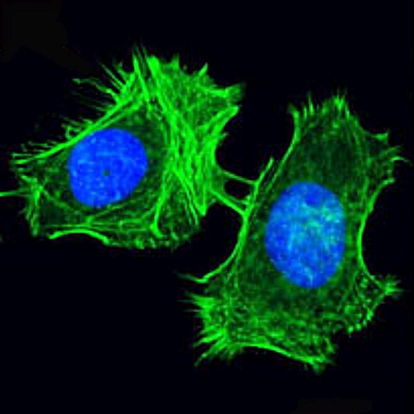
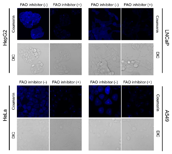
Four cancer cell lines (HepG2, LNCaP, HeLa and A549) were treated with FAOBlue in HBS buffer with or without pre-treatment of etomoxir (40 uM, 3 hours), a potent FAO inhibitor. After FAOBlue incubation, blue fluorescence (Ex. 405 nm/ Em. 430-480 nm) was observed. All cell lines showed blue fluorescence in cytosol, but pre-treatment of etomoxir clearly decreased fluorescent intensities. These results indicated the blue fluorescence was derived from FAO activity in the cells.
* Experimental condition:
HepG2, 5 uM FAOBlue for 30 min.
LNCaP, 20 uM FAOBlue for 120 min HeLa, 20 ?M FAOBlue for 120 min A549, 5 ?M FAOBlue for 30 min |
|
 |
|
| Perturbation of FAO activity by drugs | |
|
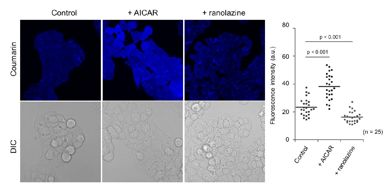
HepG2 cells were pre-incubated with 200 uM AICAR, a FAO activator via AMPK activation, for 3 hours or 200 uM of ranolazine, a partial FAO inhibitor, for 12 hours. After drug treatment, the cells were incubated with 5 uM FAOBlue for 30 min. Compared with control cells, pretreatment with AICAR significantly increased blue fluorescent intensity. On the other hand, pretreatment with the partial FAO inhibitor ranolazine clearly decreased blue fluorescent intensity. Ex. 405 nm/ Em. 430-480 nm
|
|
 |
|
| Quantitative analysis of the drug effects on FAO activity | |
|
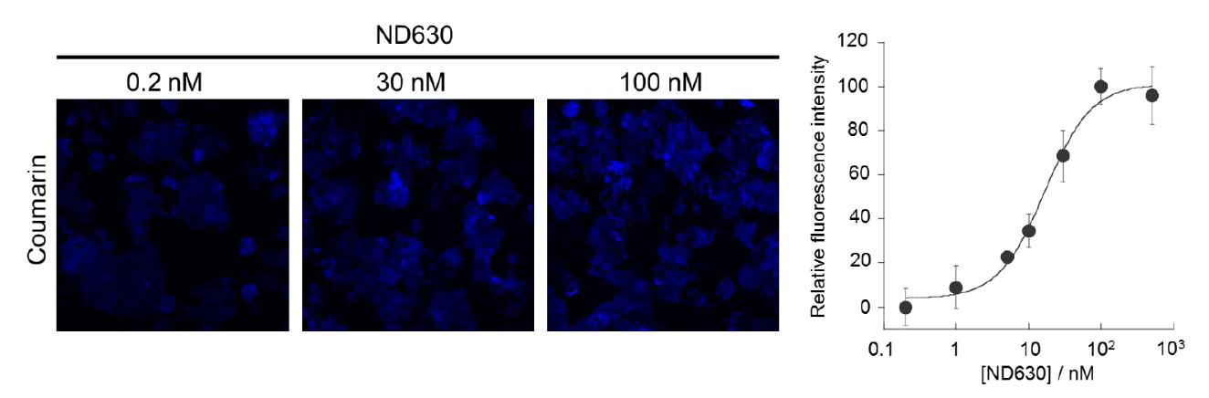
ND630 is an inhibitor of acetyl-CoA carboxylase and considered as a potential therapeutic drug for non?alcoholic fatty liver disease (NAFLD). HepG2 cells were pre-incubated with various concentration of ND630 for 4 hours. After ND630 treatment, cells were treated with 5 uM FAOBlue for 30 min. Blue fluorescent intensities of each concentration of ND630 were quantified. Ex. 405 nm/ Em. 430-480 nm
|
|
 |
|
| Analysis of FAO activity using NASH model mouse | |
|
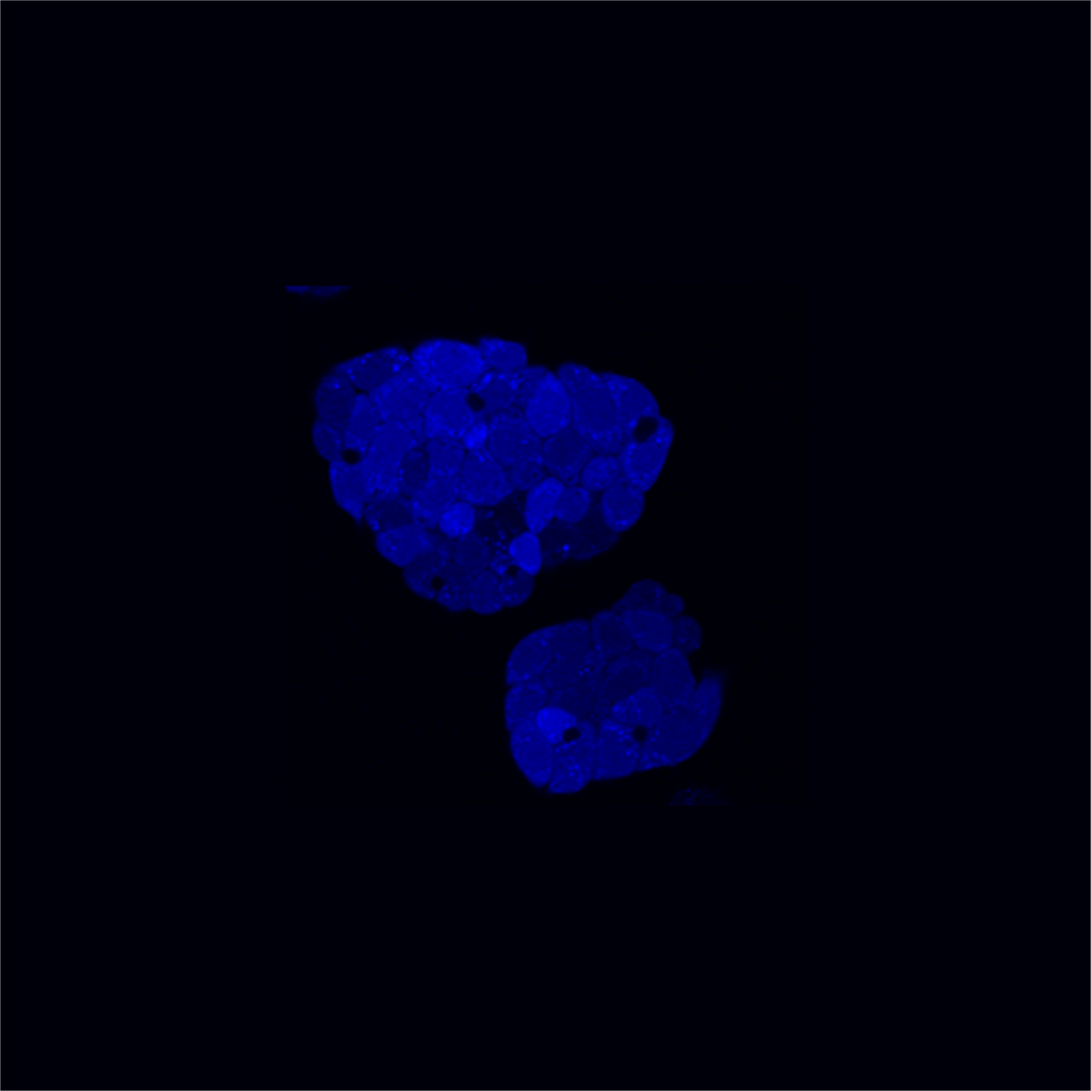
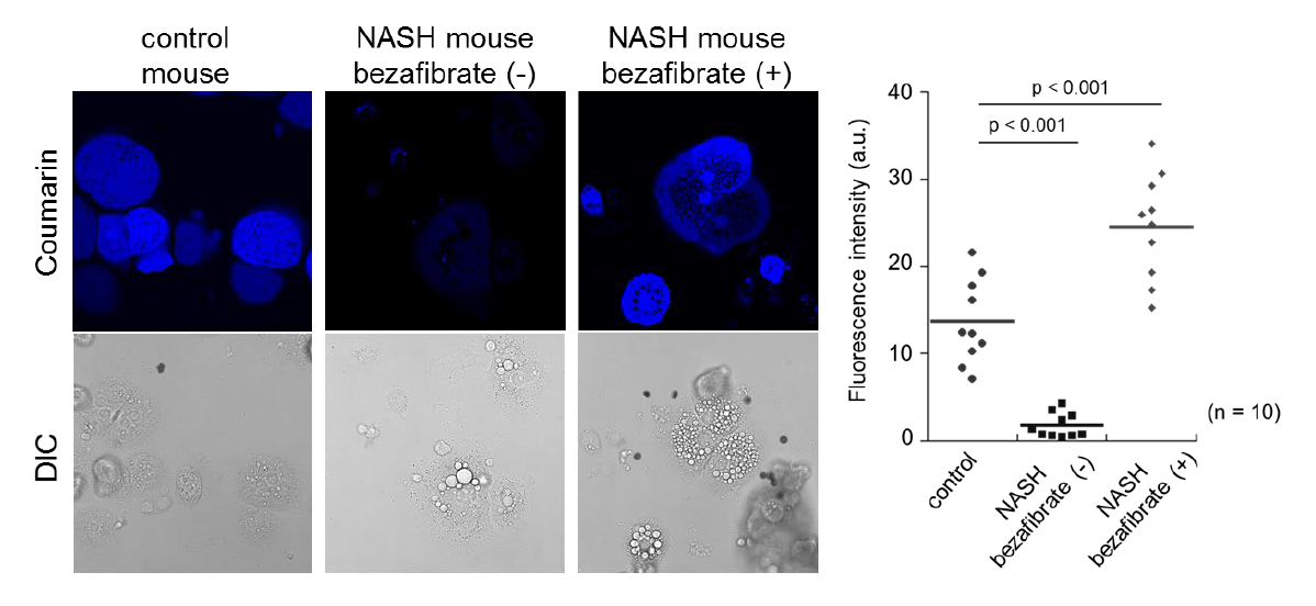
Non-alcoholic steatohepatitis (NASH) is a typical disease which shows low metabolic activity of FAs. Control healthy mice and NASH model mice were orally administered with 400 mg/kg bezafibrate, a therapeutic agent of NASH. After 4 weeks administration, primary hepatocytes were isolated from control mice and NASH model mice and cultured in culture dishes. Primary hepatocytes were further treated with 5 uM FAOBlue for 30 min and fluorescence imaging was performed. Compared with control cells, NASH model mouse-derived hepatocytes showed low FAO activity. Bezafibrate dramatically recovered FAO activity of hepatocytes isolated from NASH model mouse. FAOBlue is a powerful tool to estimate drug effects and efficiency on FAO activity. *Detail procedure of mice experiment is described in Ref.1. Ex. 405 nm/ Em. 430-480 nm |
|
 |
|
 |
We are pleased to announce that the product article about FAOBlue was posted on BioTechniques which is one of the famous international journal regarding life sciences since September 1. |
The short-chain fatty acid acetate modulates epithelial-to-mesenchymal transition
https://doi.org/10.1091/mbc.E22-02-0066
PDF file Download >>> click here <<<
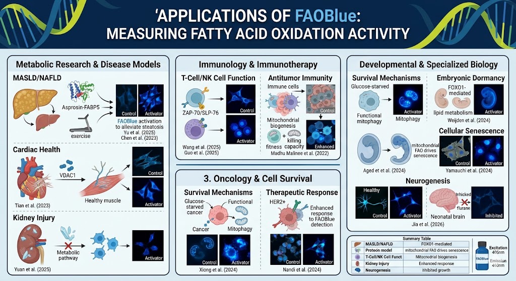
Based on the extensive list of publications provided, FAOBlue has emerged as a versatile and critical tool for directly measuring Fatty Acid Oxidation (FAO) activity across diverse biological systems.
Unlike traditional indirect methods (like oxygen consumption or radioisotopes), FAOBlue allows for real-time, fluorescence-based visualization of mitochondrial $\beta$-oxidation.
1. Metabolic Research & Disease Models
The reagent is widely used to study metabolic dysfunction, particularly in the liver and heart:
-
MASLD/NAFLD: Research by Yu et al. (2025) and Chen et al. (2023) used FAOBlue to show how specific protein interactions (Asprosin-FABP5) or exercise-induced pathways (Cdo1-AMPK) modulate fatty acid breakdown to alleviate liver steatosis.
-
Cardiac Health: Tian et al. (2023) utilized it to demonstrate how VDAC1 overexpression alleviates cardiac fibrosis by restoring healthy fatty acid metabolism.
-
Kidney Injury: Yuan et al. (2025) identified that inhibiting FAO via specific methylation pathways exacerbates crystal-induced kidney damage.
2. Immunology & Immunotherapy
FAOBlue has become a standard for assessing the metabolic state of immune cells, which often shift their energy source during activation:
-
T-Cell & NK Cell Function: It was used to show that enhancing FAO can alleviate autoimmunity in T-cells (Masuyama et al., 2024) and is involved in NK cell activation pathways (Wang et al., 2025).
-
Antitumor Immunity: Madhu Malinee et al. (2022) used the reagent to prove that mitochondrial biogenesis enhances the "fitness" and killing capacity of immune cells in tumor models.
3. Oncology & Cell Survival
Cancer cells often rewire their metabolism to survive harsh conditions:
-
Survival Mechanisms: Xiong et al. (2024) demonstrated that FAOBlue-detected activity is vital for breast cancer cells to survive glucose starvation.
-
Therapeutic Response: Nandi et al. (2024) showed that targeting FAO can actually enhance the effectiveness of HER2-targeted therapies.
4. Developmental & Specialized Biology
The reagent has expanded into niche areas of biological timing and stress:
-
Embryonic Dormancy: Used to show that lipid metabolism maintains mammalian embryos in a state of suspended animation (diapause) (Weijden et al., 2024).
-
Cellular Senescence: Yamauchi et al. (2024) discovered that mitochondrial fatty acid oxidation is a primary driver of the aging process (senescence) in cells.
- Neurogenesis: Jia et al. (2026) utilized it to show how certain anesthetics (Sevoflurane) impair cognitive function by inhibiting FAO in neural stem cells.
Uchinomiya et al., Chem. Commun., 56, 3023-3026 (2020) Fluorescence Detection of Metabolic Activity of Fatty Acid Beta Oxidation Activity in Living Cells.
Lyu et al., Molecular Biology of the Cell (2022) The short-chain fatty acid acetate modulates epithelial-to-mesenchymal transition. https://doi.org/10.1091/mbc.E22-02-0066
Shang et al., Aging (Albany NY) (2022) Systemic lipolysis promotes physiological fitness in Drosophila melanogaster. 14(16): 6481–6506.
Matsumoto A er al., (2023) Spatiotemporally quantitative in vivo imaging of mitochondrial fatty acid β-oxidation at cellular-level resolution in mice. Am J Physiol Endocrinol Metab. 325(5):E552-E561. doi: 10.1152/ajpendo.00147.
Madhu Malinee M. et al., (2022) Targeted epigenetic induction of mitochondrial biogenesis enhances antitumor immunity in mouse model. Cell Chem Biol. 17;29(3):463-475.e6. doi: 10.1016/j.chembiol.2021.08.001.
Suzuki K. et al., (2023) CLPX regulates mitochondrial fatty acid β-oxidation in liver cells. J Biol Chem. 299(10):105210. doi: 10.1016/j.jbc.2023.105210.
Tian G. et al., (2023) Voltage-dependent anion channel 1 (VDAC1) overexpression alleviates cardiac fibroblast activation in cardiac fibrosis via regulating fatty acid metabolism. Redox Biol. 67:102907. doi: 10.1016/j.redox.2023.102907.
Ikizawa T. et al., (2023) Mitochondria directly sense osmotic stress to trigger rapid metabolic remodeling via regulation of pyruvate dehydrogenase phosphorylation. J Biol Chem. 299(2):102837. doi: 10.1016/j.jbc.2022.102837.
Weijden V. et al., (2024) FOXO1-mediated lipid metabolism maintains mammalian embryos in dormancy. Nat Cell Biol. 26(2):181-193. doi: 10.1038/s41556-023-01325-3.
Chen M.et (2023) Cdo1-Camkk2-AMPK axis confers the protective effects of exercise against NAFLD in mice. Nat Commun
. 2023 Dec 18;14(1):8391. doi: 10.1038/s41467-023-44242-7.
Watanuki S., et al., (2024) Context-dependent modification of PFKFB3 in hematopoietic stem cells promotes anaerobic glycolysis and ensures stress hematopoiesis. Elife 12:RP87674.
Luo J., (2024) Dual-specificity phosphatase 5-mediated fatty acid oxidation promotes Mycobacterium bovis BCG -induced inflammatory responses. Exp Cell Res. 434(2):113869.
Nandi I., (2024) Targeting fatty acid oxidation enhances response to HER2-targeted therapy. Nat Commun. 15(1):6587.
Xiong Z et al., (2024) MANF facilitates breast cancer cell survival under glucose-starvation conditions via PRKN-mediated mitophagy regulation Autophagy. 4:1-22.
Masuyama S., et al. (2024) Enhanced fatty acid oxidation by selective activation of PPARα alleviates autoimmunity through metabolic transformation in T-cells. Clin Immunol. 268:110357.
Roy-Dorval A., et al., (2024) Analysis of lipid uptake, storage, and fatty acid oxidation by group 2 innate lymphoid cells. Front Immunol. 15:1493848
Cheng J., et al., (2024) FOXO1 induced fatty acid oxidation in hepatic cells by targeting ALDH1L2. J Gastroenterol Hepatol. 39(10):2197-2207
Lin S., et al., (2025) SNRK modulates mTOR-autophagy pathway for liver lipid homeostasis in MAFLD. Mol. Ther. 33(1):279-296
Jacobs CF., et al., (2025) Cholesterol homeostasis and lipid raft dynamics at the basis of tumor-induced immune dysfunction in chronic lymphocytic leukemia. Cell Mol Immunol. doi: 10.1038/s41423-025-01262-1
Nirgude S., et al., (2025) Single-nucleus multiomic analysis of Beckwith-Wiedemann syndrome liver reveals PPARA signaling enrichment and metabolic dysfunction. Commun Biol. 8(1):495.
EL. Braverman., et al., (2024) AMPK agonism optimizes the in vivo persistence and anti-leukemia efficacy of chimeric antigen receptor T cells. bioRxiv.2024.09.26.615290
Yuan T., et al., (2025) PRMT1-mediated methylation of UBE2m promoting calcium oxalate crystal-induced kidney injury by inhibiting fatty acid metabolism. Cell Death Dis. 2025 Jul 31;16(1):579
Seo J., et al., (2025) RNA binding protein HuD regulates fatty acid oxidation in pancreatic β-cells by modulating long-chain acyl-CoA dehydrogenase expression. Anim Cells Syst (Seoul). 2025 Aug 11;29(1):512–522.
Chen X., (2024) PAK3 Exacerbates Cardiac Lipotoxicity via SREBP1c in Obesity Cardiomyopathy. Diabetes, 73(11), 1805-1820. https://doi.org/10.2337/db24-0240
Huang, Yimin et al. Cell Host & Microbe, Volume 33, Issue 9, 1484 - 1501.e12. https://doi.org/10.1016/j.chom.2025.07.012
Zhang, C., Yang, D., Suzuki, H., Chen, J., Wang, J., Ye, M., ... & Zeng, X. (2025). Hepatocyte-specific CLSTN3B ablation impairs lipid droplet maturation and alleviates diet-induced steatohepatitis in mice. bioRxiv, 2025.05.20.655199. (Preprint)
Zhang, C., Yang, D., Suzuki, H., do Vale, G. D., Chen, J., Vaidya, A., ... & Zeng, X. (2026). Interorganelle competition for linoleic acid underlies steatotic liver pathology. bioRxiv, 2026.01.11.698890. (Preprint).
Koda, K., Kamogashira, T., Hayashi, K., Fujimoto, C., Iwasaki, S., Yamasoba, T., & Kondo, K. (2025). Rates of Mitochondrial Metabolism of Glucose, Amino Acids, and Fatty Acids by the HEI-OC1 Inner Ear Cell Line. Biology, 14(9), 1118. https://doi.org/10.3390/biology14091118
Phetkong, C., Boonto, T., Thamjamrassri, P., Ariyachet, C., & Tangkijvanich, P. (2025). MicroRNA-372-3p impairs fatty acid metabolism in hepatocellular carcinoma cells by targeting CPT1A and ACSL4. BioImpacts : BI, 15, 31075. https://doi.org/10.34172/bi.31075
Jia, P., Wang, K., Cheng, Y., Zhang, Y., Lu, Y., Sun, H., Zhang, S., Fan, P., Zhang, Y., Yang, L., Wang, N., Lu, H., Chen, X., Liu, Y., Wei, H., & Zhang, P. (2026). Sevoflurane impairs neurogenesis and cognitive function by inhibiting fatty acid β-oxidation in neural stem/progenitor cells of neonatal rats. Chinese medical journal, 139(2), 282–295. https://doi.org/10.1097/CM9.0000000000003864
Zhao, X., Liu, L., Chu, X., Zhang, Y., Tang, Y., Shao, J., Fan, B., Yang, Y., & Xu, B. (2025). Cellular osmoregulation enhances porcine embryo development by restoring zygotic genome activation via metabolic-epigenetic crosstalk. Cellular & molecular biology letters, 30(1), 137. https://doi.org/10.1186/s11658-025-00826-3
Yu, Y. Y., Feng, M., Chen, Y., Jia, H. L., Zhang, Q., Tong, M., Li, Y. X., Zhao, Y., Liu, X. X., Cao, S. F., Wang, Z. K., Li, H. W., Liu, X., & Zhang, Y. (2025). Asprosin-FABP5 Interaction Modulates Mitochondrial Fatty Acid Oxidation through PPARα Contributing to MASLD Development. Advanced science (Weinheim, Baden-Wurttemberg, Germany), 12(21), e2415846. https://doi.org/10.1002/advs.202415846
Liu, Y., Nakayama, Y., Sugita, J., Oshima, T., Kani, K., Kobayashi, A., Setoguchi, N., Iwai, Y., Manabe, I., & Fujiu, K. (2026). MALAT1 regulates human macrophage metabolism by interacting with HADHB. iScience, 29(3), 115107. https://doi.org/10.1016/j.isci.2026.115107
Cai, J., Li, W., Wu, C., Ni, H., Ran, H., Huang, Y., Tang, X., He, W., Gu, Y., You, Y., Li, J., Xiao, X., & Ma, L. (2025). β-Hydroxybutyrate enhances malate dehydrogenase 2 β-hydroxybutyrylation to alleviate hepatic steatosis in MASLD. Cellular and molecular life sciences : CMLS, 82(1), 361. https://doi.org/10.1007/s00018-025-05819-1
Shimizu, H., Horibata, Y., Amano, I., Ritter, M. J., Domae, M., Ando, H., Sugimoto, H., Cohen, R. N., & Hollenberg, A. N. (2022). Nuclear corepressor SMRT acts as a strong regulator of both β-oxidation and suppressor of fibrosis in the differentiation process of mouse skeletal muscle cells. PloS one, 17(12), e0277830. https://doi.org/10.1371/journal.pone.0277830
Si, W. Y., Yang, C. L., Wei, S. L., Du, T., Li, L. K., Dong, J., Zhou, Y., Li, H., Zhang, P., Liu, Q. J., Duan, R. S., & Duan, R. N. (2024). Therapeutic potential of microglial SMEK1 in regulating H3K9 lactylation in cerebral ischemia-reperfusion. Communications biology, 7(1), 1701. https://doi.org/10.1038/s42003-024-07425-6
Yamauchi, S., Sugiura, Y., Yamaguchi, J., Zhou, X., Takenaka, S., Odawara, T., Fukaya, S., Fujisawa, T., Naguro, I., Uchiyama, Y., Takahashi, A., & Ichijo, H. (2024). Mitochondrial fatty acid oxidation drives senescence. Science advances, 10(43), eado5887. https://doi.org/10.1126/sciadv.ado5887
Huang, N., Fang, Y., Zheng, S., Wu, J., Huang, G., Deng, X., Guan, Y., Liang, B., Zeng, D., Xu, B., Huang, Y., Rong, X., Wang, C., Bin, J., Liao, Y., Wu, Z., Shi, M., & Liao, W. (2025). Residual tumor cells after insufficient radiofrequency ablation promote lung metastasis by educating CD177hiPAD4hi neutrophils. Nature communications, 17(1), 208. https://doi.org/10.1038/s41467-025-66897-0
Flow Cytometry
Zhu, Y., Zhou, Q., Chen, Y., Zhang, Y., Wang, S., Wang, Y., ... & Wang, Y. (2025). A large-scale single-cell transcriptomic atlas of mouse brain aging. Communications Biology, 8(1), Article 427.
--- Paper citation material --- updated Apr 17, 2025 ---
Product Citation: FAO Blue (Fatty Acid Oxidation Detection Reagent)
1. LILRA3 triggers NK cell activation and cytotoxicity via ZAP-70/SLP-76 signaling pathway
Authors: Wang, W. et al.
Journal: Cellular & Molecular Immunology (Nature Publishing Group)
Publication Year: 2025
Product Mentioned: FNK-FDV-0033
Summary: This study investigates the role of LILRA3 in human NK cell activation, where FNK-FDV-0033 was used to assess cytotoxicity and signaling pathways. The results demonstrate that LILRA3 promotes NK cell cytotoxicity through the ZAP-70/SLP-76 axis.
2. Proteolysis of transcription factor ETV6 by MALT1 promotes IL-17 production in ILC3s
Authors: Guo, Q. et al.
Journal: Communications Biology (Nature Publishing Group)
Publication Year: 2025
Product Mentioned: FNK-FDV-0033
Summary: In this article, researchers uncover a mechanism by which MALT1 regulates IL-17 production in innate lymphoid cells (ILC3s). FNK-FDV-0033 was employed in the functional assays to characterize cellular immune responses and validate findings related to ETV6 cleavage.