Anti-HCV Core protein antibody, monoclonal, (FITC conjugated)
Anti-Hepatitis C Virus (HCV) core protein antibody
DiagnoCine offers excellent HCV I Hepatitis C Virus Antibodies and Reagents to researchers studying RNA Replication, Viral Structural Proteins, Transcriptional Regulation, Immunopathogenesis of HCV, HCV-induced Autophagy, Diagnostic Markers for Hepatitis, Hepatic Stellate Cells, HCV Genome, Antiviral Drug Resistance, Propagation of HCV, and other Pathogenic Factor for Hepatitis research topics.
Human diseases include Liver Cancer, Fibrosis, Fibromyalgia, Cirrhosis, Hepatocellular carcinoma, and other Chronic Liver infections & Diseases.
Anti-HCV I Hepatitis C Virus Antibodies have excellent quality and this highly pure antibody can be adapted for Western blotting, Immunofluorescence staining, Immunohistochemistry, and ELISA research with optimization
Order Information
| Cat. No. | Size | Labeled | Data Link | Link |
| FNK-65-052-S | 100 µg | Unlabeled (Clone: H6-29) |
UniProtKB P26662 |
 |
| FNK-65-052 | 100 µg | Unlabeled (Clone: H6-29) |
UniProtKB P26662 |
|
| FNK-65-053 | 50 µg | Biotinylated | Swiss-Prot HCV protein |
|
| FNK-65-054 | 50 µg | FITC-conjugated | Swiss-Prot HCV protein |
|
General information
| Cat. No. | :Table Below |
| Size | :Table Below |
| Form | :65-052: Purified monoclonal antibody (IgG) 1mg/ml in PBS, 50% glycerol, filter-sterilized :65-053: 0.7 mg/ml in PBS, 50% glycerol, filter-sterilized :65-054: 1.6 mg/ml in PBS, 50% glycerol, filter-sterilized |
| Immunogen | :A part of the core region (nucleotides 369-704, amino acids 13-124) of HCV genotype 1b expressed in E. coli (the nucleotide sequence is shown in ref.3) |
| Clone No. | :H6-29 |
| Host Animal | :Mouse |
| Isotype | :Mouse IgG2a kappa |
| Reactivity | :Reacts with human HCV core antigen, p21, of genotype 1b. Not tested in other genotypes |
| Application | :1. Western blotting (1/1,000~1/2,000) :2. Immunohistochemistry (1/100~1/500) :3. Immunofluorescence staining (1/100~1/500) :4. ELISA (assay dependent) |
| Shipping and Storage | :Ship at 4℃ and store at -20℃. Do not freeze. |
Description
Hepatitis C virus (HCV) is a small (55-65 nm in size), enveloped, positive sense single-stranded RNA virus in the family Flaviviridae and the principal cause of parenteral non-A, non-B hepatitis. The virus genome consists of a single open reading frame of approximately 9,4 kb which encodes a single polyprotein of about 3,010 amino acids (1, 2, 3). The polyprotein is processed by host cell and viral proteases into four structural proteins (core, envelope1 and 2, and p7) and six non-structural proteins (NS2, 3, 4a, 4b, 5a, and 5b) necessary for viral replication. HCV core protein is not only a component of nucleocapsid but also has multiple functions and is thought to be a pathogenic factor for hepatitis. It also participates in some cellular processes, including transcriptional regulation and cellular transduction. HCV core antigen is used as diagnostic marker for HCV infection.
Figures
 |
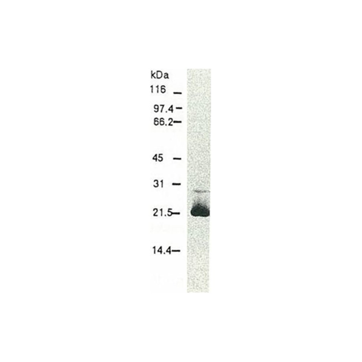
Fig.1 Western blotting of HCV core protein. Chimp liver cells were infected with recombinant vaccinia virus containing a HCV genome cDNA and were subjected to Western blotting using this antibody. The core protein is detected as a 22-kDa band. | |
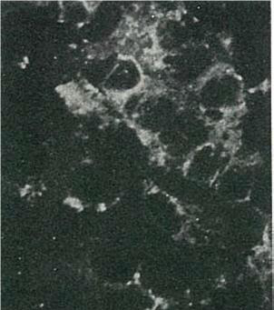 |
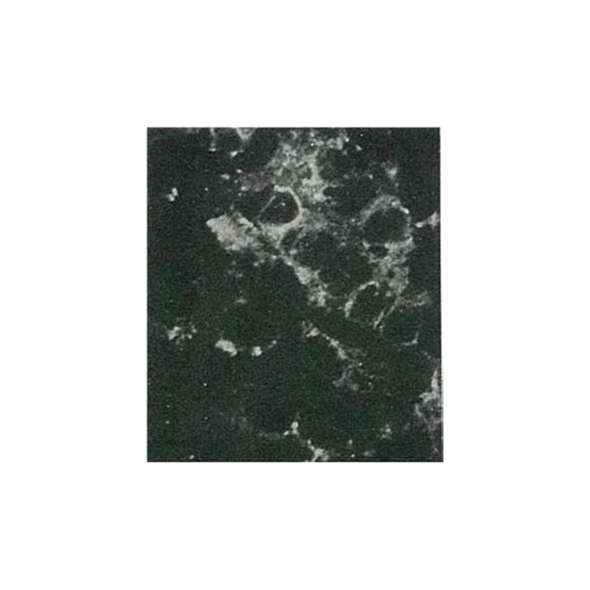
Fig.2 Detection of HCV core protein by immunofluorescence antibody staining. Chimp liver cells were infected with recombinant vaccinia virus containing a HCV genome cDNA. After incubation for 48 hr, the cells were fixed with acetone and HCV core protein was detected by indirect immunofluorescence staining using this antibody | |
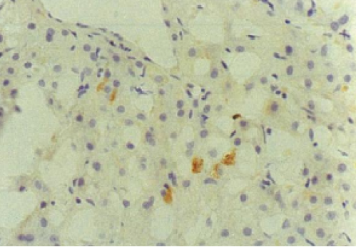 |
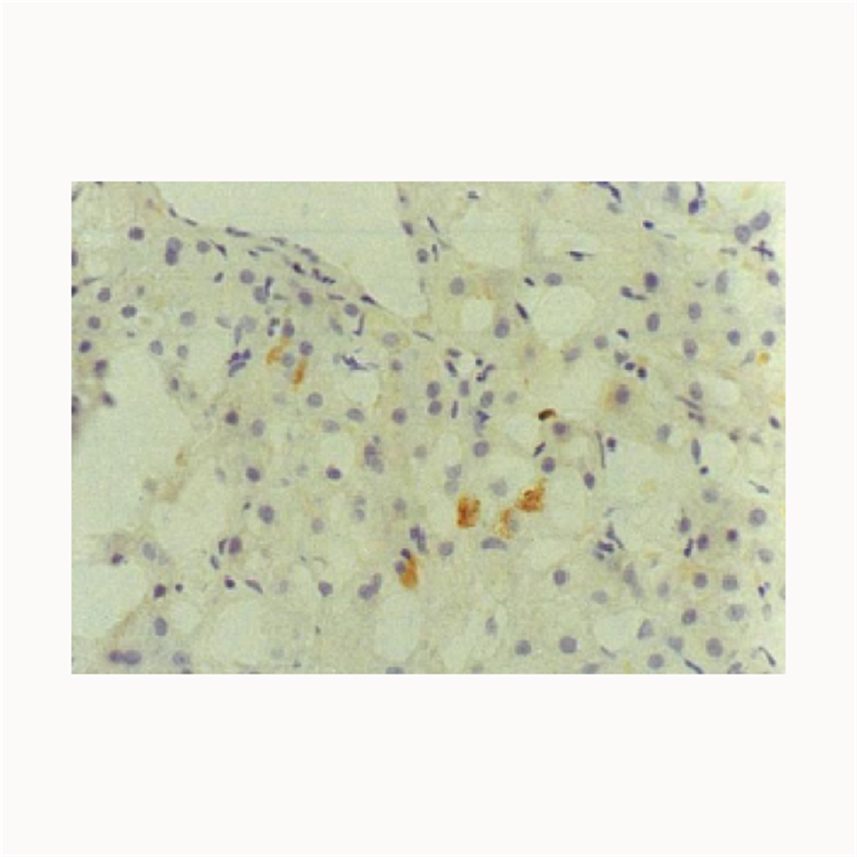
Fig.3 Immunohistochemical detection of HCV core protein. Tissue section from a patient with chronic hepatitis C was immunostained to reveal cells expressing HCV core antigen, which are scattered in the lobules (indirect immunohistochemical method, counterstained with Mayer’s hematoxylin). |
References
- Brass V, Moradpour D, Blum HE. Molecular Virology of Hepatitis C Virus (HCV): 2006 Update. Int J Med Sci 2006; 3:29-34. PMID: 16614739
- Kato, N. et al. (1990) “Molecular cloning of the human hepatitis C virus genome from Japanese patients with non-A, non-B hepatitis.” Proc. Natl. Acad. Sci. USA 87, 9524-9528 PMID: 2175903
- Takamizawa, A. et al. (1991) “Structure and organization of the hepatitis C virus genome isolated from human carriers.” J.Virol.65, 1105-1113 PMID: 1847440
- Manabe, S. et al. (1994) “Production of nonstructural proteins of hepatitis C virus requires a putative viral protease encoded by N3.” Virology 198, 636-644 PMID: 8291245
- Hiramatsu, N. et al. (1992) “Immunohistochemical detection of hepatitis C virus-infected hepatocytes in chronic liver disease with monoclonal antibodies to core, envelope and NS3 regions of the hepatitis C virus genome.” Hepatology, 16, 306-311 PMID: 1379209