

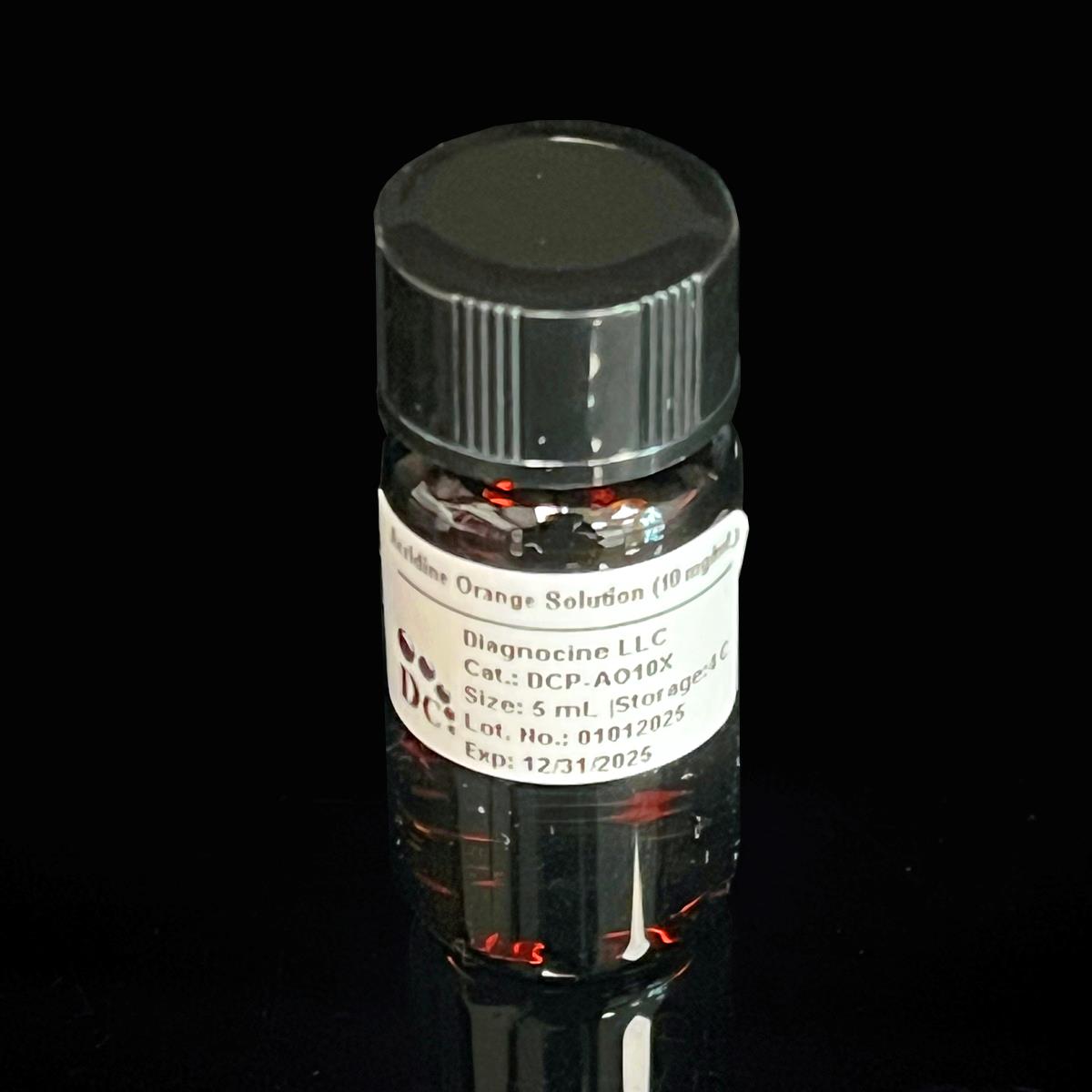
Acridine Orange Solution (10 mg/mL)
Cat No.: DCP-AO10X
Size: 5 mL
Sterile, Ultrapure, Filtered 0.22-micron membrane.
Description:This 10 mg/mL Acridine Orange staining solution is a high-purity, ready-to-use fluorescent nucleic acid dye optimized for cellular and microbiological applications. Prepared in molecular-grade water, the solution contains Acridine Orange hydrochloride (CAS 65-61-2), a metachromatic fluorophore that differentially stains DNA and RNA with distinct spectral emissions. The formulation ensures consistent performance in live-cell imaging, flow cytometry, and clinical diagnostics, with validated stability under recommended storage conditions.
Key Features:
- Dual Fluorescence Emission: Binds dsDNA to emit green fluorescence (Ex/Em: 500/526 nm) and RNA/ssDNA to emit red fluorescence (Ex/Em: 460/650 nm).
- Cell Permeability: Suitable for live and fixed cells, enabling staining of intracellular nucleic acids and acidic compartments (e.g., lysosomes).
- High Sensitivity: Detects microorganisms at concentrations as low as 10⁴ CFU/mL, ideal for rapid clinical specimen analysis.
Applications:
- Cell Cycle Analysis: Distinguishes G0/G1, S, and G2/M phases via DNA/RNA quantification in flow cytometry.
- Microorganism Detection: Rapid screening of bacteria, yeast, and acid-fast bacilli in cerebrospinal fluid, blood cultures, and tissue smears.
- Apoptosis/Necroptosis Assays: Identifies apoptotic cells (red fluorescence) and necroptotic cells (distinct spectral signatures).
- Lysosomal Tracking: Accumulates in acidic organelles for real-time lysosomal membrane permeability studies.
- Intraoperative Tumor Imaging: Guides surgical resection of gliomas and other malignancies through fluorescence contrast.
Composition:
| Ingredients | Concentration |
| Acridine Orange Hydrochloride | 10 mg/mL |
Usage Instructions:
General Guidelines:
-
Working Concentration: Dilute to 1–20 µg/mL depending on application (see below).
-
Light Protection: Perform all steps in low-light conditions to prevent photobleaching.
-
Controls: Include unstained cells and RNase-treated samples (for RNA specificity).
1. Flow Cytometry (Cell Cycle/Apoptosis Analysis)
Materials:
-
Cells in suspension (1 × 10⁶/mL in PBS)
-
Acridine Orange working solution (1 µg/mL in PBS)
-
Flow cytometer with 488 nm excitation and 530/640 nm emission filters
Steps:
-
Prepare Cells: Wash cells twice with PBS and resuspend in PBS at 1 × 10⁶ cells/mL.
-
Dilute Stain: Add 1 µL of 10 mg/mL stock to 10 mL PBS (1 µg/mL final concentration).
-
Stain Cells: Incubate cells with working solution for 15 min at 37°C.
-
Analyze: Acquire data immediately using a flow cytometer. Gate populations based on green (DNA) vs. red (RNA) fluorescence.
Notes:
-
For apoptosis assays, compare fluorescence ratios (red/green) between treated and untreated cells.
-
Avoid prolonged incubation (>30 min) to prevent cytotoxicity.
2. Fluorescence Microscopy (Microorganism/Cell Staining)
Materials:
-
Fixed or live cells on slides
-
Methanol (for fixation)
-
Fluorescence microscope with blue (460–490 nm) excitation filter
Steps:
-
Fixation: Fix cells with ice-cold methanol for 2 min. Air-dry.
-
Stain: Apply 100 µL of diluted AO (5 µg/mL in PBS, pH 4.0) for 2 min.
-
Rinse: Wash slides 3× with PBS (pH 4.0) to remove unbound dye.
-
Mount: Add coverslip with PBS or antifade mounting medium.
-
Image: Visualize using a 40×–100× objective.
-
DNA: Green fluorescence (FITC filter).
-
RNA/Acidic organelles: Red fluorescence (TRITC filter).
-
Notes:
-
For live-cell imaging, reduce dye concentration to 1–2 µg/mL to minimize toxicity.
-
Bacterial cells stain bright orange; human cells appear pale green.
3. Acid-Fast Bacilli (AFB) Staining
Materials:
-
Sputum/tissue smears
-
Phenol-based decolorizer (e.g., 3% HCl in ethanol)
Steps:
-
Heat-Fix Smear: Pass slide through a flame 3× after air-drying.
-
Stain: Flood slide with AO working solution (10 µg/mL in 1% phenol) for 15 min.
-
Decolorize: Rinse with 1% HCl in ethanol for 2 min.
-
Counterstain (Optional): Apply 0.1% potassium permanganate for 30 sec.
-
Visualize: Examine under fluorescence microscope (200× magnification).
-
AFB: Bright orange-yellow rods against a dark background.
-
4. Lysosomal Staining (Live Cells)
Steps:
-
Prepare Working Solution: Dilute stock to 5 µg/mL in cell culture medium.
-
Incubate: Add to cells and incubate for 10–15 min at 37°C.
-
Wash: Replace medium with dye-free buffer.
-
Image: Use time-lapse microscopy to track lysosomal pH changes (red fluorescence).
- PPE: Gloves, lab coat, and eye protection required.
- Waste Disposal: Collect in hazardous waste containers; AO is a potential mutagen.
- Appearance: Yellow-orange solution
- Purity: ≥95% (HPLC), zinc-free
- Sterility: Filtered 0.22 micron.
- DNase activity: None was detected after incubation of plasmid DNA, and this product was used for 18 hours at room temperature.
- RNase activity: No RNase activity was detected after incubation of ribosomal RNA and this product for 18 hr at room temperature.
Storage and Shelf Life:
- Store at 4°C away from bright light.
- Shelf life is 12 months.
- Use before expiry date given on the product label.
CUSTOMIZATION: Please INQUIRE if other concentrations, additions of chemicals/compounds/proteins/supplements, different pH, and modifications are needed.
All DiagnoCine Precision Sterile buffers are filtered-sterilized with a 0.1-micron filtration two times and 0.04-micron filtration one time. Thus, mycoplasma contamination is prevented. The smallest size mycoplasma type can be about 0.2 microns.
* This product is manufactured under ISO 13485-certified and CE-approved facilities (Suppliers of DiagnoCine Precision). All final packaging, quality assurance, and testing are done at the DiagnoCine R&D and Quality Testing Center. All specific customization requests and assembly were accomplished at DiagnoCine Precision in Totowa, New Jersey, USA.